5HCJ | pdb_00005hcj

Cationic Ligand-Gated Ion Channel


Experimental Data Snapshot

  • Method: X-RAY DIFFRACTION
  • Resolution: 2.95 Å
  • R-Value Free: 
    0.218 (Depositor), 0.239 (DCC) 
  • R-Value Work: 
    0.213 (Depositor), 0.234 (DCC) 
  • R-Value Observed: 
    0.214 (Depositor) 

wwPDB Validation 3D Report Full Report

Validation slider image for 5HCJ

Ligand Structure Quality Assessment 


This is version 1.2 of the entry. See complete history

Literature

Sites of Anesthetic Inhibitory Action on a Cationic Ligand-Gated Ion Channel.

Laurent, B.Murail, S.Shahsavar, A.Sauguet, L.Delarue, M.Baaden, M.

(2016) Structure 24: 595-605

  • DOI: https://doi.org/10.1016/j.str.2016.02.014
  • Primary Citation Related Structures: 
    5HCJ, 5HCM

  • PubMed Abstract: 

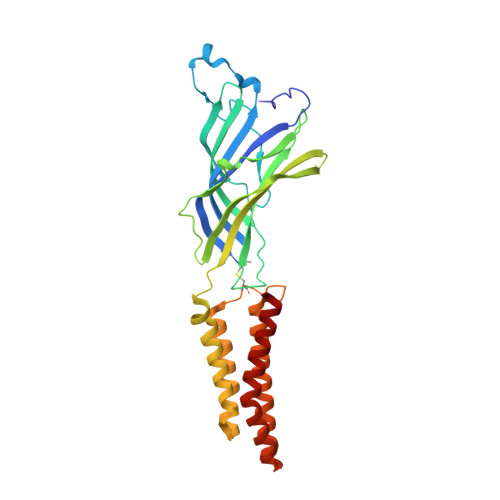
    Pentameric ligand-gated ion channels have been identified as the principal target of general anesthetics (GA), whose molecular mechanism of action remains poorly understood. Bacterial homologs, such as the Gloeobacter violaceus receptor (GLIC), have been shown to be valid functional models of GA action. The GA bromoform inhibits GLIC at submillimolar concentration. We characterize bromoform binding by crystallography and molecular dynamics (MD) simulations. GLIC's open form structure identified three intra-subunit binding sites. We crystallized the locally closed form with an additional bromoform molecule in the channel pore. We systematically compare binding with the multiple potential sites of allosteric channel regulation in the open, locally closed, and resting forms. MD simulations reveal differential exchange pathways between sites from one form to the other. GAs predominantly access the receptor from the lipid bilayer in all cases. Differential binding affinity among the channel forms is observed; the pore site markedly stabilizes the inactive versus active state.


  • Organizational Affiliation
    • Laboratoire de Biochimie Théorique, CNRS, UPR9080, University Paris Diderot, Sorbonne Paris Cité, 13 rue Pierre et Marie Curie, 75005 Paris, France.

Macromolecule Content 

  • Total Structure Weight: 182.16 kDa 
  • Atom Count: 12,743 
  • Modeled Residue Count: 1,555 
  • Deposited Residue Count: 1,585 
  • Unique protein chains: 1

Macromolecules

Find similar proteins by:|  3D Structure
Entity ID: 1
MoleculeChains  Sequence LengthOrganismDetailsImage
Proton-gated ion channel
A, B, C, D, E
317Gloeobacter violaceusMutation(s): 3 
Gene Names: glvIglr4197
Membrane Entity: Yes 
UniProt
Find proteins for Q7NDN8 (Gloeobacter violaceus (strain ATCC 29082 / PCC 7421))
Explore Q7NDN8 
Go to UniProtKB:  Q7NDN8
Entity Groups
Sequence Clusters30% Identity50% Identity70% Identity90% Identity95% Identity100% Identity
UniProt GroupQ7NDN8
Sequence Annotations
Expand
Reference Sequence

Small Molecules

Ligands 4 Unique
IDChains Name / Formula / InChI Key2D Diagram3D Interactions
LMT

Query on LMT


Download:Ideal Coordinates CCD File
F [auth A]DODECYL-BETA-D-MALTOSIDE
C24 H46 O11
NLEBIOOXCVAHBD-QKMCSOCLSA-N
MBR

Query on MBR


Download:Ideal Coordinates CCD File
M [auth E]TRIBROMOMETHANE
C H Br3
DIKBFYAXUHHXCS-UHFFFAOYSA-N
CL

Query on CL


Download:Ideal Coordinates CCD File
G [auth A],
I [auth B],
J [auth C],
K [auth D],
L [auth E]
CHLORIDE ION
Cl
VEXZGXHMUGYJMC-UHFFFAOYSA-M
NA

Query on NA


Download:Ideal Coordinates CCD File
H [auth A]SODIUM ION
Na
FKNQFGJONOIPTF-UHFFFAOYSA-N

Experimental Data & Validation

Experimental Data

  • Method: X-RAY DIFFRACTION
  • Resolution: 2.95 Å
  • R-Value Free:  0.218 (Depositor), 0.239 (DCC) 
  • R-Value Work:  0.213 (Depositor), 0.234 (DCC) 
  • R-Value Observed: 0.214 (Depositor) 
Space Group: C 1 2 1
Unit Cell:
Length ( Å )Angle ( ˚ )
a = 181.944α = 90
b = 133.372β = 102.34
c = 159.337γ = 90
Software Package:
Software NamePurpose
BUSTERrefinement
XDSdata reduction
SCALAdata scaling

Structure Validation

View Full Validation Report


Ligand Structure Quality Assessment 


Entry History 

Deposition Data

Revision History  (Full details and data files)

  • Version 1.0: 2016-04-13
    Type: Initial release
  • Version 1.1: 2016-04-20
    Changes: Database references
  • Version 1.2: 2024-11-13
    Changes: Data collection, Database references, Structure summary