


Crystal structure of NADH bound carbonyl reductase from Streptomyces coelicolor
Li, M., Kong, X.-D., Zhou, J., Yu, H.-L., Zhang, Z.-J., Xu, J.-H.To be published.
Experimental Data Snapshot
Entity ID: 1 | |||||
|---|---|---|---|---|---|
| Molecule | Chains | Sequence Length | Organism | Details | Image |
| Putative oxidoreductase | 263 | Streptomyces coelicolor A3(2) | Mutation(s): 0 Gene Names: SCO7363 | 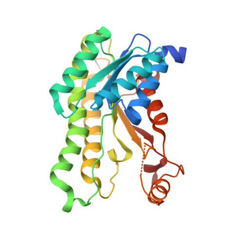 | |
UniProt | |||||
Entity Groups | |||||
| Sequence Clusters | 30% Identity50% Identity70% Identity90% Identity95% Identity100% Identity | ||||
| UniProt Group | Q9KYM4 | ||||
Sequence AnnotationsExpand | |||||
Reference Sequence | |||||
| Ligands 3 Unique | |||||
|---|---|---|---|---|---|
| ID | Chains | Name / Formula / InChI Key | 2D Diagram | 3D Interactions | |
| NAI Download:Ideal Coordinates CCD File | DA [auth F] GA [auth G] JA [auth H] M [auth A] NA [auth I] | 1,4-DIHYDRONICOTINAMIDE ADENINE DINUCLEOTIDE C21 H29 N7 O14 P2 BOPGDPNILDQYTO-NNYOXOHSSA-N |  | ||
| IPA Download:Ideal Coordinates CCD File | BA [auth E] CA [auth E] EA [auth F] FA [auth F] HA [auth G] | ISOPROPYL ALCOHOL C3 H8 O KFZMGEQAYNKOFK-UHFFFAOYSA-N |  | ||
| MG Download:Ideal Coordinates CCD File | AA [auth E] KA [auth H] OA [auth I] U [auth C] XA [auth L] | MAGNESIUM ION Mg JLVVSXFLKOJNIY-UHFFFAOYSA-N |  | ||
| Length ( Å ) | Angle ( ˚ ) |
|---|---|
| a = 187.787 | α = 90 |
| b = 187.787 | β = 90 |
| c = 80.858 | γ = 120 |
| Software Name | Purpose |
|---|---|
| PHENIX | refinement |
| HKL-2000 | data collection |
| HKL-2000 | data scaling |
| PHASER | phasing |
| PDB_EXTRACT | data extraction |
| HKL-2000 | data reduction |
| PHASER | phasing |
| HKL | data reduction |
| HKL | data scaling |


