Studies on the regulatory mechanism of isocitrate dehydrogenase 2 using acetylation mimics
Xu, Y., Liu, L., Nakamura, A., Someya, S., Miyakawa, T., Tanokura, M.(2017) Sci Rep 7: 9785-9785
- PubMed: 28852116 Search on PubMedSearch on PubMed Central
- DOI: https://doi.org/10.1038/s41598-017-10337-7
- Primary Citation Related Structures:
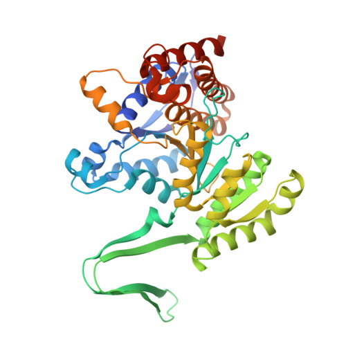
5H3E, 5H3F - PubMed Abstract:
Mitochondrial isocitrate dehydrogenase 2 (IDH2) converts NADP + to NADPH and promotes regeneration of reduced glutathione (GSH) by supplying NADPH to glutathione reductase or thioredoxin reductase. We have previously shown that under calorie restriction, mitochondrial deacetylase Sirt3 deacetylates and activates IDH2, thereby regulating the mitochondrial glutathione antioxidant defense system in mice. To investigate the regulatory mechanism of mIDH2 (mouse mitochondrial IDH2), we used lysine-to-glutamine (KQ) mutants to mimic acetylated lysines and screened 15 KQ mutants. Among these mutants, the activities of the K256Q and K413Q proteins were less than 50% of the wild-type value. We then solved the crystal structures of the wild-type mIDH2 and the K256Q mutant proteins, revealing conformational changes in the substrate-binding pocket. Structural data suggested that positively charged Lys256 was important in stabilizing the pocket because it repelled a lysine cluster on the other side. Glutamine (or acetylated lysine) was neutral and thus caused the pocket size to decrease, which might be the main reason for the lower activity of the K256Q mutant. Together, our data provide the first structure of an acetylation mimic of mIDH2 and new insights into the regulatory mechanism of acetylation of mIDH2.
- Laboratory of Basic Science on Healthy Longevity, Department of Applied Biological Chemistry, Graduate School of Agricultural and Life Sciences, The University of Tokyo, 1-1-1 Yayoi, Bunkyo-ku, Tokyo, 113-8657, Japan.
Organizational Affiliation: