5H2O | pdb_00005h2o

A three dimensional movie of structural changes in bacteriorhodopsin: structure obtained 250 us after photoexcitation


Experimental Data Snapshot

  • Method: X-RAY DIFFRACTION
  • Resolution: 2.10 Å
  • R-Value Free: 
    0.184 (Depositor), 0.189 (DCC) 
  • R-Value Work: 
    0.161 (Depositor), 0.166 (DCC) 
  • R-Value Observed: 
    0.162 (Depositor) 

Starting Model: experimental
View more details

wwPDB Validation 3D Report Full Report

Validation slider image for 5H2O

Ligand Structure Quality Assessment 


This is version 1.5 of the entry. See complete history

Literature

A three-dimensional movie of structural changes in bacteriorhodopsin

Nango, E.Royant, A.Kubo, M.Nakane, T.Wickstrand, C.Kimura, T.Tanaka, T.Tono, K.Song, C.Tanaka, R.Arima, T.Yamashita, A.Kobayashi, J.Hosaka, T.Mizohata, E.Nogly, P.Sugahara, M.Nam, D.Nomura, T.Shimamura, T.Im, D.Fujiwara, T.Yamanaka, Y.Jeon, B.Nishizawa, T.Oda, K.Fukuda, M.Andersson, R.Bath, P.Dods, R.Davidsson, J.Matsuoka, S.Kawatake, S.Murata, M.Nureki, O.Owada, S.Kameshima, T.Hatsui, T.Joti, Y.Schertler, G.Yabashi, M.Bondar, A.N.Standfuss, J.Neutze, R.Iwata, S.

(2016) Science 354: 1552-1557

  • DOI: https://doi.org/10.1126/science.aah3497
  • Primary Citation Related Structures: 
    5B6V, 5B6W, 5B6X, 5B6Y, 5B6Z, 5H2H, 5H2I, 5H2J, 5H2K, 5H2L, 5H2M, 5H2N, 5H2O, 5H2P

  • PubMed Abstract: 

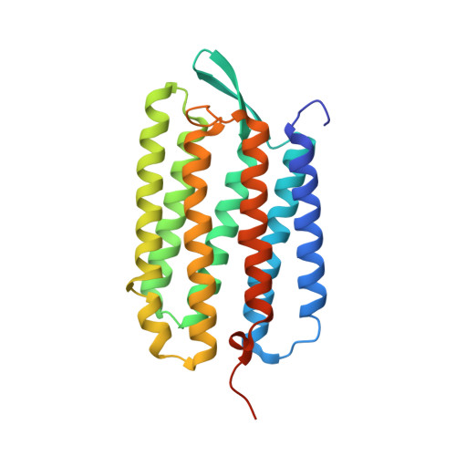
    Bacteriorhodopsin (bR) is a light-driven proton pump and a model membrane transport protein. We used time-resolved serial femtosecond crystallography at an x-ray free electron laser to visualize conformational changes in bR from nanoseconds to milliseconds following photoactivation. An initially twisted retinal chromophore displaces a conserved tryptophan residue of transmembrane helix F on the cytoplasmic side of the protein while dislodging a key water molecule on the extracellular side. The resulting cascade of structural changes throughout the protein shows how motions are choreographed as bR transports protons uphill against a transmembrane concentration gradient.


  • Organizational Affiliation
    • RIKEN SPring-8 Center, 1-1-1 Kouto, Sayo-cho, Sayo-gun, Hyogo 679-5148, Japan.

Macromolecule Content 

  • Total Structure Weight: 30.89 kDa 
  • Atom Count: 2,562 
  • Modeled Residue Count: 231 
  • Deposited Residue Count: 248 
  • Unique protein chains: 1

Macromolecules

Find similar proteins by:|  3D Structure
Entity ID: 1
MoleculeChains  Sequence LengthOrganismDetailsImage
Bacteriorhodopsin248Halobacterium salinarum NRC-1Mutation(s): 0 
Membrane Entity: Yes 
UniProt
Find proteins for P02945 (Halobacterium salinarum (strain ATCC 700922 / JCM 11081 / NRC-1))
Explore P02945 
Go to UniProtKB:  P02945
Entity Groups
Sequence Clusters30% Identity50% Identity70% Identity90% Identity95% Identity100% Identity
UniProt GroupP02945
Sequence Annotations
Expand
Reference Sequence

Small Molecules

Ligands 10 Unique
IDChains Name / Formula / InChI Key2D Diagram3D Interactions
L2P

Query on L2P


Download:Ideal Coordinates CCD File
C [auth A],
K [auth A],
L [auth A]
2,3-DI-PHYTANYL-GLYCEROL
C43 H88 O3
ISDBCJSGCHUHFI-UMZPFTBHSA-N
RET

Query on RET


Download:Ideal Coordinates CCD File
B [auth A]RETINAL
C20 H28 O
NCYCYZXNIZJOKI-OVSJKPMPSA-N
MYS

Query on MYS


Download:Ideal Coordinates CCD File
I [auth A],
M [auth A]
PENTADECANE
C15 H32
YCOZIPAWZNQLMR-UHFFFAOYSA-N
C14

Query on C14


Download:Ideal Coordinates CCD File
O [auth A]TETRADECANE
C14 H30
BGHCVCJVXZWKCC-UHFFFAOYSA-N
TRD

Query on TRD


Download:Ideal Coordinates CCD File
D [auth A]TRIDECANE
C13 H28
IIYFAKIEWZDVMP-UHFFFAOYSA-N
UND

Query on UND


Download:Ideal Coordinates CCD File
J [auth A],
Q [auth A]
UNDECANE
C11 H24
RSJKGSCJYJTIGS-UHFFFAOYSA-N
D10

Query on D10


Download:Ideal Coordinates CCD File
E [auth A]DECANE
C10 H22
DIOQZVSQGTUSAI-UHFFFAOYSA-N
DD9

Query on DD9


Download:Ideal Coordinates CCD File
N [auth A]nonane
C9 H20
BKIMMITUMNQMOS-UHFFFAOYSA-N
OCT

Query on OCT


Download:Ideal Coordinates CCD File
G [auth A],
H [auth A],
P [auth A]
N-OCTANE
C8 H18
TVMXDCGIABBOFY-UHFFFAOYSA-N
HP6

Query on HP6


Download:Ideal Coordinates CCD File
F [auth A]HEPTANE
C7 H16
IMNFDUFMRHMDMM-UHFFFAOYSA-N

Experimental Data & Validation

Experimental Data

  • Method: X-RAY DIFFRACTION
  • Resolution: 2.10 Å
  • R-Value Free:  0.184 (Depositor), 0.189 (DCC) 
  • R-Value Work:  0.161 (Depositor), 0.166 (DCC) 
  • R-Value Observed: 0.162 (Depositor) 
Space Group: P 63
Unit Cell:
Length ( Å )Angle ( ˚ )
a = 62.5α = 90
b = 62.5β = 90
c = 112γ = 120
Software Package:
Software NamePurpose
PHENIXrefinement
CrystFELdata reduction
CrystFELdata scaling

Structure Validation

View Full Validation Report


Ligand Structure Quality Assessment 


Entry History 

Deposition Data

Revision History  (Full details and data files)

  • Version 1.0: 2016-12-21
    Type: Initial release
  • Version 1.1: 2017-02-01
    Changes: Database references
  • Version 1.2: 2018-01-31
    Changes: Data collection
  • Version 1.3: 2023-09-06
    Changes: Data collection, Database references
  • Version 1.4: 2023-11-08
    Changes: Refinement description
  • Version 1.5: 2024-11-20
    Changes: Structure summary