5GXT | pdb_00005gxt

Crystal structure of PigG


Experimental Data Snapshot

  • Method: X-RAY DIFFRACTION
  • Resolution: 2.25 Å
  • R-Value Free: 
    0.296 (Depositor), 0.295 (DCC) 
  • R-Value Work: 
    0.230 (Depositor), 0.231 (DCC) 
  • R-Value Observed: 
    0.233 (Depositor) 

wwPDB Validation 3D Report Full Report

Validation slider image for 5GXT

This is version 1.2 of the entry. See complete history

Literature

Crystal structure of MBP-PigG fusion protein and the essential function of PigG in the prodigiosin biosynthetic pathway in Serratia marcescens FS14.

Zhang, F.Wei, Q.Tong, H.Xu, D.Wang, W.Ran, T.

(2017) Int J Biol Macromol 99: 394-400

  • DOI: https://doi.org/10.1016/j.ijbiomac.2017.02.088
  • Primary Citation Related Structures: 
    5GXT, 5GXV

  • PubMed Abstract: 

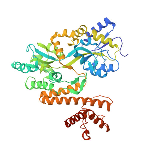
    Prodigiosin, a tripyrrole red pigment is synthesized by Serratia and some other microbes through a bifurcated biosynthesis pathway; MBC (4-methoxy-2,2'-bipyrrole-5-carbaldehyde) and MAP (2-methyl-3-n-amyl-pyrrole) are synthesized separately and then condensed by PigC to form prodigiosin. PigI, PigG and PigA have been shown to be involved in the first steps of MBC biosynthesis (proline incorporation). The crystal structure of PigG was resolved to elucidate its function and mechanism. PigG, an acyl carrier protein (ACP), features the ACP architecture:, a helical bundle fold containing three major helices and a minor distorted helix together with a conserved "S" motif. An in-frame deletion mutation of the pigG gene abolished the synthesis of prodigiosin in Serratia marcescens FS14. The production of prodigiosin was fully restored by complementation of intact pigG; however the S36A mutant was not able to restore function in the in-frame deletion pigG mutant, indicating that PigG and the conserved serine residue (S36) of PigG are essential for the synthesis of prodigiosin.


  • Organizational Affiliation
    • Key Laboratory of Microbiological Engineering of Agricultural Environment, Ministry of Agriculture, Department of Microbiology, College of Life Sciences, Nanjing Agricultural University, Nanjing, China.

Macromolecule Content 

  • Total Structure Weight: 51.42 kDa 
  • Atom Count: 3,593 
  • Modeled Residue Count: 454 
  • Deposited Residue Count: 468 
  • Unique protein chains: 1

Macromolecules

Find similar proteins by:|  3D Structure
Entity ID: 1
MoleculeChains  Sequence LengthOrganismDetailsImage
Maltose-binding periplasmic protein,PigG468Escherichia coli K-12Serratia sp. FS14
This entity is chimeric
Mutation(s): 0 
Gene Names: malEb4034JW3994
UniProt
Find proteins for A0ACD6BAW2 (Serratia sp. (strain FS14))
Explore A0ACD6BAW2 
Go to UniProtKB:  A0ACD6BAW2
Entity Groups
Sequence Clusters30% Identity50% Identity70% Identity90% Identity95% Identity100% Identity
UniProt GroupA0ACD6BAW2
Sequence Annotations
Expand
Reference Sequence

Small Molecules

Ligands 1 Unique
IDChains Name / Formula / InChI Key2D Diagram3D Interactions
MG

Query on MG


Download:Ideal Coordinates CCD File
B [auth A]MAGNESIUM ION
Mg
JLVVSXFLKOJNIY-UHFFFAOYSA-N

Experimental Data & Validation

Experimental Data

  • Method: X-RAY DIFFRACTION
  • Resolution: 2.25 Å
  • R-Value Free:  0.296 (Depositor), 0.295 (DCC) 
  • R-Value Work:  0.230 (Depositor), 0.231 (DCC) 
  • R-Value Observed: 0.233 (Depositor) 
Space Group: C 1 2 1
Unit Cell:
Length ( Å )Angle ( ˚ )
a = 95.7α = 90
b = 60β = 99.3
c = 81.4γ = 90
Software Package:
Software NamePurpose
PHENIXrefinement
XDSdata reduction
XSCALEdata scaling
PHASERphasing

Structure Validation

View Full Validation Report


Entry History 

& Funding Information

Deposition Data


Funding OrganizationLocationGrant Number
National Natural Science Foundation of ChinaChina31400055
National Natural Science Foundation of ChinaChina31170686
National Natural Science Foundation of ChinaChina31100028
the Natural Science Foundation of Jiangsu ProvinceChinaBK20140690

Revision History  (Full details and data files)

  • Version 1.0: 2017-07-19
    Type: Initial release
  • Version 1.1: 2017-10-18
    Changes: Author supporting evidence
  • Version 1.2: 2024-03-20
    Changes: Data collection, Database references