


Structure of acryloyl-CoA lyase PrpE from Dinoroseobacter shibae DFL 12
Zhang, Y.Z., Wang, P., Cao, H.Y.To be published.
Experimental Data Snapshot
Starting Model: experimental
View more details
Entity ID: 1 | |||||
|---|---|---|---|---|---|
| Molecule | Chains | Sequence Length | Organism | Details | Image |
| AMP-dependent synthetase and ligase | 630 | Dinoroseobacter shibae DFL 12 = DSM 16493 | Mutation(s): 0 Gene Names: Dshi_0825 EC: 6.2.1 | 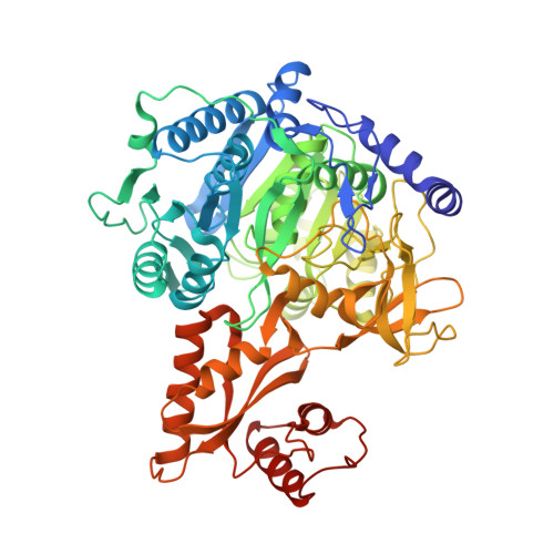 | |
UniProt | |||||
Entity Groups | |||||
| Sequence Clusters | 30% Identity50% Identity70% Identity90% Identity95% Identity100% Identity | ||||
| UniProt Group | A8LRC0 | ||||
Sequence AnnotationsExpand | |||||
Reference Sequence | |||||
| Ligands 4 Unique | |||||
|---|---|---|---|---|---|
| ID | Chains | Name / Formula / InChI Key | 2D Diagram | 3D Interactions | |
| COA Download:Ideal Coordinates CCD File | D [auth A] | COENZYME A C21 H36 N7 O16 P3 S RGJOEKWQDUBAIZ-IBOSZNHHSA-N |  | ||
| AMP Download:Ideal Coordinates CCD File | B [auth A] | ADENOSINE MONOPHOSPHATE C10 H14 N5 O7 P UDMBCSSLTHHNCD-KQYNXXCUSA-N |  | ||
| GOL Download:Ideal Coordinates CCD File | E [auth A], F [auth A], G [auth A], H [auth A], I [auth A] | GLYCEROL C3 H8 O3 PEDCQBHIVMGVHV-UHFFFAOYSA-N |  | ||
| AKR Download:Ideal Coordinates CCD File | C [auth A] | ACRYLIC ACID C3 H4 O2 NIXOWILDQLNWCW-UHFFFAOYSA-N |  | ||
| Length ( Å ) | Angle ( ˚ ) |
|---|---|
| a = 147.097 | α = 90 |
| b = 58.425 | β = 128.45 |
| c = 113.259 | γ = 90 |
| Software Name | Purpose |
|---|---|
| PHENIX | refinement |
| HKL-2000 | data reduction |
| HKL-2000 | data scaling |
| PHASER | phasing |
| Funding Organization | Location | Grant Number |
|---|---|---|
| China | -- |


