5GUE

Crystal structure of CotB2 (GGSPP/Mg2+-Bound Form) from Streptomyces melanosporofaciens


Experimental Data Snapshot

  • Method: X-RAY DIFFRACTION
  • Resolution: 1.80 Å
  • R-Value Free: 0.231 
  • R-Value Work: 0.191 
  • R-Value Observed: 0.193 

wwPDB Validation   3D Report Full Report


Ligand Structure Quality Assessment 


This is version 1.2 of the entry. See complete history


Literature

Structural Insights into the CotB2-Catalyzed Cyclization of Geranylgeranyl Diphosphate to the Diterpene Cyclooctat-9-en-7-ol

Tomita, T.Kim, S.-Y.Teramoto, K.Meguro, A.Ozaki, T.Yoshida, A.Motoyoshi, Y.Mori, N.Ishigami, K.Watanabe, H.Nishiyama, M.Kuzuyama, T.

(2017) ACS Chem Biol 12: 1621-1628

  • DOI: https://doi.org/10.1021/acschembio.7b00154
  • Primary Citation of Related Structures:  
    5GUC, 5GUE

  • PubMed Abstract: 

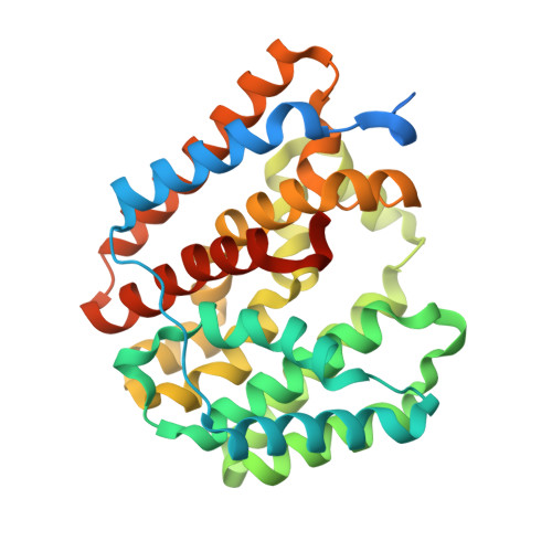
    The diterpene cyclase CotB2 catalyzes the cyclization of geranylgeranyl diphosphate (GGPP) to the tricyclic cyclooctat-9-en-7-ol, which is characterized by a 5-8-5-fused ring skeleton. We have previously proposed a cyclization cascade involving a unique carbon-carbon bond rearrangement combined with multiple hydride shifts, all occurring at a single active site. Here, we report the first high-resolution X-ray crystal structure of CotB2 with bound substrate analog geranylgeranyl thiodiphosphate (GGSPP). In the GGSPP-bound form, GGSPP folds into a unique S-shaped conformation that probably reflects the substrate-bound state prior to ionization of the substrate GGPP. The folded framework of GGSPP is surrounded by hydrophobic residues and several aromatic and asparagine residues that are well-positioned to stabilize a series of reactive carbocation intermediates through a combination of cation-π and dipole charge interactions. The combined crystal structures and mutagenesis-based biochemical assays provide a structural basis for exquisite control of ring formation and stereochemistry during CotB2 catalysis.


  • Organizational Affiliation

    RIKEN SPring-8 Center , 1-1-1 Kouto, Sayo-cho, Sayo-gun, Hyogo 679-5148, Japan.


Macromolecules
Find similar proteins by:  (by identity cutoff)  |  3D Structure
Entity ID: 1
MoleculeChains Sequence LengthOrganismDetailsImage
Cyclooctat-9-en-7-ol synthase
A, B, C, D
331Streptomyces melanosporofaciensMutation(s): 0 
Gene Names: CotB2
EC: 4.2.3.146
UniProt
Find proteins for C9K1X5 (Streptomyces melanosporofaciens)
Explore C9K1X5 
Go to UniProtKB:  C9K1X5
Entity Groups  
Sequence Clusters30% Identity50% Identity70% Identity90% Identity95% Identity100% Identity
UniProt GroupC9K1X5
Sequence Annotations
Expand
  • Reference Sequence
Experimental Data & Validation

Experimental Data

  • Method: X-RAY DIFFRACTION
  • Resolution: 1.80 Å
  • R-Value Free: 0.231 
  • R-Value Work: 0.191 
  • R-Value Observed: 0.193 
  • Space Group: P 1 21 1
Unit Cell:
Length ( Å )Angle ( ˚ )
a = 58.698α = 90
b = 100.373β = 90
c = 108.937γ = 90
Software Package:
Software NamePurpose
HKL-2000data reduction
HKL-2000data scaling
PHASERphasing
REFMACrefinement

Structure Validation

View Full Validation Report


Ligand Structure Quality Assessment 


Entry History & Funding Information

Deposition Data


Funding OrganizationLocationGrant Number
JSPSJapan26292058

Revision History  (Full details and data files)

  • Version 1.0: 2017-06-07
    Type: Initial release
  • Version 1.1: 2017-07-05
    Changes: Database references
  • Version 1.2: 2023-11-08
    Changes: Data collection, Database references, Derived calculations, Refinement description