5GTD | pdb_00005gtd

o-Succinylbenzoate CoA Synthetase (MenE) from Bacillus Subtilis in Complex with the Acyl-adenylate Intermediate OSB-AMP


Experimental Data Snapshot

  • Method: X-RAY DIFFRACTION
  • Resolution: 2.69 Å
  • R-Value Free: 
    0.237 (Depositor), 0.276 (DCC) 
  • R-Value Work: 
    0.200 (Depositor) 
  • R-Value Observed: 
    0.202 (Depositor) 

Starting Model: experimental
View more details

wwPDB Validation 3D Report Full Report

Validation slider image for 5GTD

Ligand Structure Quality Assessment 


This is version 2.0 of the entry. See complete history

Literature

Mechanistic Insights from the Crystal Structure of Bacillus subtilis o-Succinylbenzoyl-CoA Synthetase Complexed with the Adenylate Intermediate

Chen, Y.Jiang, Y.Guo, Z.

(2016) Biochemistry 55: 6685-6695

  • DOI: https://doi.org/10.1021/acs.biochem.6b00889
  • Primary Citation Related Structures: 
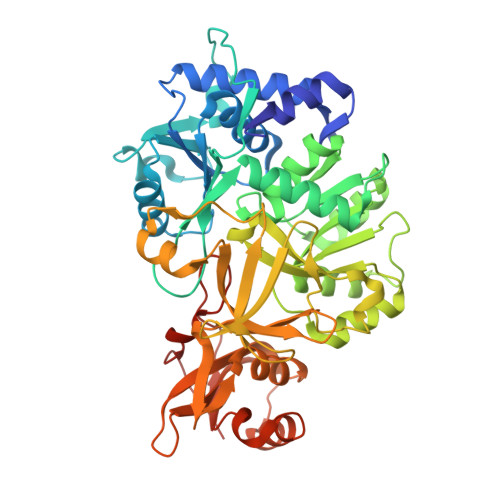
    5GTD

  • PubMed Abstract: 

    o-Succinylbenzoyl-CoA (OSB-CoA) synthetase, or MenE, catalyzes an essential step in vitamin K biosynthesis and is a valuable drug target. Like many other adenylating enzymes, it changes its structure to accommodate substrate binding, catalysis, and product release along the path of a domain alternation catalytic mechanism. We have determined the crystal structure of its complex with the adenylation product, o-succinylbenzoyl-adenosine monophosphate (OSB-AMP), and captured a new postadenylation state. This structure presents unique features such as a strained conformation for the bound adenylate intermediate to indicate that it represents the enzyme state after completion of the adenylation reaction but before release of the C domain in its transition to the thioesterification conformation. By comparison to the ATP-bound preadenylation conformation, structural changes are identified in both the reactants and the active site to allow inference about how these changes accommodate and facilitate the adenylation reaction and to directly support an in-line backside attack nucleophilic substitution mechanism for the first half-reaction. Mutational analysis suggests that the conserved His196 plays an important role in desolvation of the active site rather than stabilizing the transition state of the adenylation reaction. In addition, comparison of the new structure with a previously determined OSB-AMP-bound structure of the same enzyme allows us to propose a release mechanism of the C domain in its alteration to form the thioesterification conformation. These findings allow us to better understand the domain alternation catalytic mechanism of MenE as well as many other adenylating enzymes.


  • Organizational Affiliation
    • Department of Chemistry, The Hong Kong University of Science and Technology , Clear Water Bay, Kowloon, Hong Kong SAR, China.

Macromolecule Content 

  • Total Structure Weight: 112.02 kDa 
  • Atom Count: 6,495 
  • Modeled Residue Count: 864 
  • Deposited Residue Count: 988 
  • Unique protein chains: 1

Macromolecules

Find similar proteins by:|  3D Structure
Entity ID: 1
MoleculeChains  Sequence LengthOrganismDetailsImage
2-succinylbenzoate--CoA ligase
A, B
494Bacillus subtilis subsp. subtilis str. 168Mutation(s): 0 
Gene Names: menEBSU30790
EC: 6.2.1.26
UniProt
Find proteins for P23971 (Bacillus subtilis (strain 168))
Explore P23971 
Go to UniProtKB:  P23971
Entity Groups
Sequence Clusters30% Identity50% Identity70% Identity90% Identity95% Identity100% Identity
UniProt GroupP23971
Sequence Annotations
Expand
Reference Sequence

Small Molecules

Ligands 6 Unique
IDChains Name / Formula / InChI Key2D Diagram3D Interactions
AMP

Query on AMP


Download:Ideal Coordinates CCD File
G [auth A],
L [auth B]
ADENOSINE MONOPHOSPHATE
C10 H14 N5 O7 P
UDMBCSSLTHHNCD-KQYNXXCUSA-N
OSB

Query on OSB


Download:Ideal Coordinates CCD File
H [auth A],
M [auth B]
2-SUCCINYLBENZOATE
C11 H10 O5
YIVWQNVQRXFZJB-UHFFFAOYSA-N
PEG

Query on PEG


Download:Ideal Coordinates CCD File
F [auth A]DI(HYDROXYETHYL)ETHER
C4 H10 O3
MTHSVFCYNBDYFN-UHFFFAOYSA-N
IMD

Query on IMD


Download:Ideal Coordinates CCD File
K [auth B]IMIDAZOLE
C3 H5 N2
RAXXELZNTBOGNW-UHFFFAOYSA-O
CL

Query on CL


Download:Ideal Coordinates CCD File
C [auth A],
D [auth A],
E [auth A],
I [auth B]
CHLORIDE ION
Cl
VEXZGXHMUGYJMC-UHFFFAOYSA-M
MG

Query on MG


Download:Ideal Coordinates CCD File
J [auth B]MAGNESIUM ION
Mg
JLVVSXFLKOJNIY-UHFFFAOYSA-N

Experimental Data & Validation

Experimental Data

  • Method: X-RAY DIFFRACTION
  • Resolution: 2.69 Å
  • R-Value Free:  0.237 (Depositor), 0.276 (DCC) 
  • R-Value Work:  0.200 (Depositor) 
  • R-Value Observed: 0.202 (Depositor) 
Space Group: P 43
Unit Cell:
Length ( Å )Angle ( ˚ )
a = 121.814α = 90
b = 121.814β = 90
c = 98.563γ = 90
Software Package:
Software NamePurpose
PHENIXrefinement
HKL-2000data reduction
HKL-2000data scaling
PHENIXphasing

Structure Validation

View Full Validation Report


Ligand Structure Quality Assessment 


Entry History 

& Funding Information

Deposition Data


Funding OrganizationLocationGrant Number
the University Grants Council of the Government of the Hong Kong Special Administrative RegionHong KongSBI14SC05

Revision History  (Full details and data files)

  • Version 1.0: 2017-01-04
    Type: Initial release
  • Version 1.1: 2023-11-08
    Changes: Data collection, Database references, Derived calculations, Refinement description
  • Version 2.0: 2024-05-29
    Changes: Advisory, Atomic model, Data collection, Derived calculations, Non-polymer description, Structure summary