5GMY | pdb_00005gmy

Crystal structure of the Archaeoglobus fulgidus oligosaccharyltransferase (O29867_ARCFU) tethered with an acceptor peptide containing the NVT sequon via a disulfide bond


Experimental Data Snapshot

  • Method: X-RAY DIFFRACTION
  • Resolution: 3.50 Å
  • R-Value Free: 
    0.274 (Depositor), 0.279 (DCC) 
  • R-Value Work: 
    0.226 (Depositor), 0.231 (DCC) 
  • R-Value Observed: 
    0.228 (Depositor) 

Starting Model: experimental
View more details

wwPDB Validation 3D Report Full Report

Validation slider image for 5GMY

This is version 2.1 of the entry. See complete history

Literature

Tethering an N-Glycosylation Sequon-Containing Peptide Creates a Catalytically Competent Oligosaccharyltransferase Complex

Matsumoto, S.Taguchi, Y.Shimada, A.Igura, M.Kohda, D.

(2017) Biochemistry 56: 602-611

  • DOI: https://doi.org/10.1021/acs.biochem.6b01089
  • Primary Citation Related Structures: 
    5GMY

  • PubMed Abstract: 

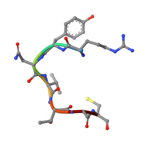
    Oligosaccharyltransferase (OST) transfers an oligosaccharide chain to the Asn residue in the Asn-X-Ser/Thr sequon in proteins, where X is not proline. A sequon was tethered to an archaeal OST enzyme via a disulfide bond. The positions of the cysteine residues in the OST protein and the sequon-containing acceptor peptide were selected by reference to the eubacterial OST structure in a noncovalent complex with an acceptor peptide. We determined the crystal structure of the cross-linked OST-sequon complex. The Ser/Thr-binding pocket recognizes the Thr residue in the sequon, and the catalytic structure termed the "carboxylate dyad" interacted with the Asn residue. Thus, the recognition and the catalytic mechanism of the sequon are conserved between the archaeal and eubacterial OSTs. We found that the tethered peptides in the complex were efficiently glycosylated in the presence of the oligosaccharide donor. The stringent requirements are greatly relaxed in the cross-linked state. The two conserved acidic residues in the catalytic structure were each dispensable, although the double mutation abolished the activity. A Gln residue at the Asn position in the sequon functioned as an acceptor, and the hydroxy group at position +2 was not required. In the standard assay using short free peptides, strong amino acid preferences were observed at the X position, but the preferences, except for Pro, completely disappeared in the cross-linked state. By skipping the initial binding process and stabilizing the complex state, the catalytically competent cross-linked complex offers a unique system for studying the oligosaccharyl transfer reaction.


  • Organizational Affiliation
    • Division of Structural Biology, Medical Institute of Bioregulation, ‡Research Center for Advanced Immunology, and §Research Center for Live-Protein Dynamics, Kyushu University , Maidashi 3-1-1, Higashi-ku, Fukuoka 812-8582, Japan.

Macromolecule Content 

  • Total Structure Weight: 100.01 kDa 
  • Atom Count: 6,887 
  • Modeled Residue Count: 855 
  • Deposited Residue Count: 882 
  • Unique protein chains: 2

Macromolecules

Find similar proteins by:|  3D Structure
Entity ID: 1
MoleculeChains  Sequence LengthOrganismDetailsImage
Transmembrane oligosaccharyl transferase, putative875Archaeoglobus fulgidus DSM 4304Mutation(s): 1 
Gene Names: AF_0380
EC: 2.4.99.18 (PDB Primary Data), 2.4.99.21 (UniProt)
Membrane Entity: Yes 
UniProt
Find proteins for O29867 (Archaeoglobus fulgidus (strain ATCC 49558 / DSM 4304 / JCM 9628 / NBRC 100126 / VC-16))
Explore O29867 
Go to UniProtKB:  O29867
Entity Groups
Sequence Clusters30% Identity50% Identity70% Identity90% Identity95% Identity100% Identity
UniProt GroupO29867
Sequence Annotations
Expand
Reference Sequence
Find similar proteins by:  Sequence   |   3D Structure  
Entity ID: 2
MoleculeChains  Sequence LengthOrganismDetailsImage
acceptor peptide, ARG-TYR-ASN-VAL-THR-ALA-CYS7synthetic constructMutation(s): 0 

Small Molecules

Ligands 1 Unique
IDChains Name / Formula / InChI Key2D Diagram3D Interactions
MG

Query on MG


Download:Ideal Coordinates CCD File
C [auth A]MAGNESIUM ION
Mg
JLVVSXFLKOJNIY-UHFFFAOYSA-N

Experimental Data & Validation

Experimental Data

  • Method: X-RAY DIFFRACTION
  • Resolution: 3.50 Å
  • R-Value Free:  0.274 (Depositor), 0.279 (DCC) 
  • R-Value Work:  0.226 (Depositor), 0.231 (DCC) 
  • R-Value Observed: 0.228 (Depositor) 
Space Group: P 43 21 2
Unit Cell:
Length ( Å )Angle ( ˚ )
a = 121.549α = 90
b = 121.549β = 90
c = 181.344γ = 90
Software Package:
Software NamePurpose
PHENIXrefinement
HKL-2000data reduction
HKL-2000data scaling
PHENIXphasing

Structure Validation

View Full Validation Report


Entry History 

& Funding Information

Deposition Data


Funding OrganizationLocationGrant Number
Japan Society for the Promotion of Science (JSPS)JapanJP24370047, JP26119002

Revision History  (Full details and data files)

  • Version 1.0: 2017-02-01
    Type: Initial release
  • Version 1.1: 2017-02-15
    Changes: Database references
  • Version 1.2: 2020-02-26
    Changes: Data collection
  • Version 2.0: 2021-03-31
    Type: Coordinate replacement
    Reason: Ligand geometry
    Changes: Advisory, Atomic model, Author supporting evidence, Data collection, Database references, Derived calculations, Other, Refinement description, Source and taxonomy, Structure summary
  • Version 2.1: 2023-11-08
    Changes: Data collection, Database references, Refinement description