5GKP

Crystal structure of the EndoG worm homologue CPS-6 H148A/F122A in complex with DNA


Experimental Data Snapshot

  • Method: X-RAY DIFFRACTION
  • Resolution: 2.30 Å
  • R-Value Free: 0.253 
  • R-Value Work: 0.173 
  • R-Value Observed: 0.180 

wwPDB Validation   3D Report Full Report


This is version 1.2 of the entry. See complete history


Literature

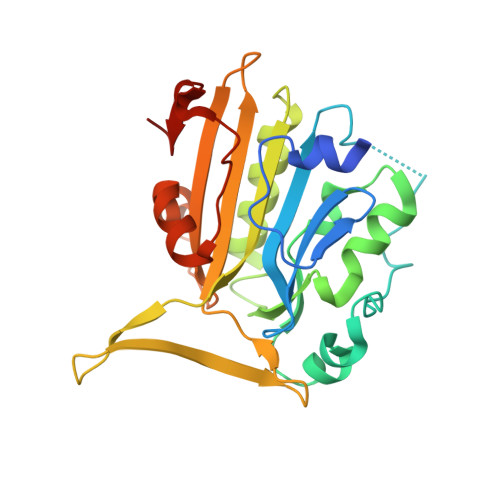
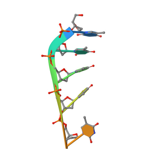
Crystal structure of endonuclease G in complex with DNA reveals how it nonspecifically degrades DNA as a homodimer.

Lin, J.L.Wu, C.C.Yang, W.Z.Yuan, H.S.

(2016) Nucleic Acids Res 44: 10480-10490

  • DOI: https://doi.org/10.1093/nar/gkw931
  • Primary Citation of Related Structures:  
    5GKC, 5GKP

  • PubMed Abstract: 

    Endonuclease G (EndoG) is an evolutionarily conserved mitochondrial protein in eukaryotes that digests nucleus chromosomal DNA during apoptosis and paternal mitochondrial DNA during embryogenesis. Under oxidative stress, homodimeric EndoG becomes oxidized and converts to monomers with diminished nuclease activity. However, it remains unclear why EndoG has to function as a homodimer in DNA degradation. Here, we report the crystal structure of the Caenorhabditis elegans EndoG homologue, CPS-6, in complex with single-stranded DNA at a resolution of 2.3 Å. Two separate DNA strands are bound at the ββα-metal motifs in the homodimer with their nucleobases pointing away from the enzyme, explaining why CPS-6 degrades DNA without sequence specificity. Two obligatory monomeric CPS-6 mutants (P207E and K131D/F132N) were constructed, and they degrade DNA with diminished activity due to poorer DNA-binding affinity as compared to wild-type CPS-6. Moreover, the P207E mutant exhibits predominantly 3'-to-5' exonuclease activity, indicating a possible endonuclease to exonuclease activity change. Thus, the dimer conformation of CPS-6 is essential for maintaining its optimal DNA-binding and endonuclease activity. Compared to other non-specific endonucleases, which are usually monomeric enzymes, EndoG is a unique dimeric endonuclease, whose activity hence can be modulated by oxidation to induce conformational changes.


  • Organizational Affiliation

    Institute of Molecular Biology, Academia Sinica, Taipei, Taiwan 11529, ROC.


Macromolecules

Find similar proteins by:  (by identity cutoff)  |  3D Structure
Entity ID: 1
MoleculeChains Sequence LengthOrganismDetailsImage
Endonuclease G, mitochondrial
A, B
252Caenorhabditis elegansMutation(s): 2 
Gene Names: cps-6C41D11.8
EC: 3.1.30
UniProt
Find proteins for Q95NM6 (Caenorhabditis elegans)
Explore Q95NM6 
Go to UniProtKB:  Q95NM6
Entity Groups  
Sequence Clusters30% Identity50% Identity70% Identity90% Identity95% Identity100% Identity
UniProt GroupQ95NM6
Sequence Annotations
Expand
  • Reference Sequence

Find similar nucleic acids by:  Sequence   |   3D Structure  

Entity ID: 2
MoleculeChains LengthOrganismImage
DNA (5'-D(*TP*TP*TP*TP*T)-3')
C, D
8unidentified
Sequence Annotations
Expand
  • Reference Sequence
Experimental Data & Validation

Experimental Data

  • Method: X-RAY DIFFRACTION
  • Resolution: 2.30 Å
  • R-Value Free: 0.253 
  • R-Value Work: 0.173 
  • R-Value Observed: 0.180 
  • Space Group: P 21 21 21
Unit Cell:
Length ( Å )Angle ( ˚ )
a = 56.672α = 90
b = 72.562β = 90
c = 127.914γ = 90
Software Package:
Software NamePurpose
PHENIXrefinement
HKL-2000data reduction
HKL-2000data scaling
PHENIXphasing

Structure Validation

View Full Validation Report


Entry History & Funding Information

Deposition Data


Funding OrganizationLocationGrant Number
Academia Sinica and the National Science Council, TaiwanTaiwan--

Revision History  (Full details and data files)

  • Version 1.0: 2016-11-23
    Type: Initial release
  • Version 1.1: 2017-12-06
    Changes: Database references, Derived calculations
  • Version 1.2: 2023-11-08
    Changes: Data collection, Database references, Derived calculations, Refinement description