Lysine Decarboxylase with an Enhanced Affinity for Pyridoxal 5-Phosphate by Disulfide Bond-Mediated Spatial Reconstitution
Sagong, H.-Y., Kim, K.-J.(2017) PLoS One 12: e0170163-e0170163
- PubMed: 28095457 Search on PubMedSearch on PubMed Central
- DOI: https://doi.org/10.1371/journal.pone.0170163
- Primary Citation Related Structures:
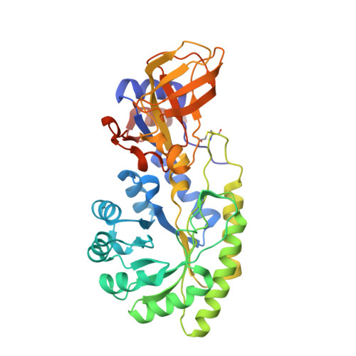
5GJO - PubMed Abstract:
Lysine decarboxylase (LDC) catalyzes the decarboxylation of l-lysine to produce cadaverine, an important industrial platform chemical for bio-based polyamides. However, due to high flexibility at the pyridoxal 5-phosphate (PLP) binding site, use of the enzyme for cadaverine production requires continuous supplement of large amounts of PLP. In order to develop an LDC enzyme from Selenomonas ruminantium (SrLDC) with an enhanced affinity for PLP, we introduced an internal disulfide bond between Ala225 and Thr302 residues with a desire to retain the PLP binding site in a closed conformation. The SrLDCA225C/T302C mutant showed a yellow color and the characteristic UV/Vis absorption peaks for enzymes with bound PLP, and exhibited three-fold enhanced PLP affinity compared with the wild-type SrLDC. The mutant also exhibited a dramatically enhanced LDC activity and cadaverine conversion particularly under no or low PLP concentrations. Moreover, introduction of the disulfide bond rendered SrLDC more resistant to high pH and temperature. The formation of the introduced disulfide bond and the maintenance of the PLP binding site in the closed conformation were confirmed by determination of the crystal structure of the mutant. This study shows that disulfide bond-mediated spatial reconstitution can be a platform technology for development of enzymes with enhanced PLP affinity.
- School of Life Sciences, KNU Creative BioResearch Group, Kyungpook National University, Daegu, Republic of Korea.
Organizational Affiliation: