5GIQ | pdb_00005giq

Xaa-Pro peptidase from Deinococcus radiodurans, Zinc bound


Experimental Data Snapshot

  • Method: X-RAY DIFFRACTION
  • Resolution: 1.80 Å
  • R-Value Free: 
    0.195 (Depositor), 0.196 (DCC) 
  • R-Value Work: 
    0.165 (Depositor), 0.165 (DCC) 
  • R-Value Observed: 
    0.166 (Depositor) 

Starting Model: experimental
View more details

wwPDB Validation 3D Report Full Report

Validation slider image for 5GIQ

This is version 1.2 of the entry. See complete history

Literature

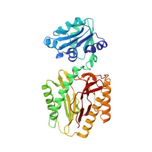
Structures and activities of widely conserved small prokaryotic aminopeptidases-P clarify classification of M24B peptidases.

Are, V.N.Kumar, A.Goyal, V.D.Gotad, S.S.Ghosh, B.Gadre, R.Jamdar, S.N.Makde, R.D.

(2018) Proteins 

  • DOI: https://doi.org/10.1002/prot.25641
  • Primary Citation Related Structures: 
    5CNX, 5GIQ

  • PubMed Abstract: 

    M24B peptidases cleaving Xaa-Pro bond in dipeptides are prolidases whereas those cleaving this bond in longer peptides are aminopeptidases-P. Bacteria have small aminopeptidases-P (36-39 kDa), which are diverged from canonical aminopeptidase-P of Escherichia coli (50 kDa). Structure-function studies of small aminopeptidases-P are lacking. We report crystal structures of small aminopeptidases-P from E. coli and Deinococcus radiodurans, and report substrate-specificities of these proteins and their ortholog from Mycobacterium tuberculosis. These are aminopeptidases-P, structurally close to small prolidases except for absence of dipeptide-selectivity loop. We noticed absence of this loop and conserved arginine in canonical archaeal prolidase (Maher et al., Biochemistry. 43, 2004, 2771-2783) and questioned its classification. Our enzymatic assays show that this enzyme is an aminopeptidase-P. Further, our mutagenesis studies illuminate importance of DXRY sequence motif in bacterial small aminopeptidases-P and suggest common evolutionary origin with human XPNPEP1/XPNPEP2. Our analyses reveal sequence/structural features distinguishing small aminopeptidases-P from other M24B peptidases.


  • Organizational Affiliation
    • High Pressure and Synchrotron Radiation Physics Division, Bhabha Atomic Research Centre, Mumbai, Maharashtra, India.

Macromolecule Content 

  • Total Structure Weight: 37.37 kDa 
  • Atom Count: 2,913 
  • Modeled Residue Count: 348 
  • Deposited Residue Count: 349 
  • Unique protein chains: 1

Macromolecules

Find similar proteins by:|  3D Structure
Entity ID: 1
MoleculeChains  Sequence LengthOrganismDetailsImage
Proline dipeptidase349Deinococcus radiodurans R1 = ATCC 13939 = DSM 20539Mutation(s): 1 
Gene Names: DR_1246
UniProt
Find proteins for Q9RUY4 (Deinococcus radiodurans (strain ATCC 13939 / DSM 20539 / JCM 16871 / CCUG 27074 / LMG 4051 / NBRC 15346 / NCIMB 9279 / VKM B-1422 / R1))
Explore Q9RUY4 
Go to UniProtKB:  Q9RUY4
Entity Groups
Sequence Clusters30% Identity50% Identity70% Identity90% Identity95% Identity100% Identity
UniProt GroupQ9RUY4
Sequence Annotations
Expand
Reference Sequence

Experimental Data & Validation

Experimental Data

  • Method: X-RAY DIFFRACTION
  • Resolution: 1.80 Å
  • R-Value Free:  0.195 (Depositor), 0.196 (DCC) 
  • R-Value Work:  0.165 (Depositor), 0.165 (DCC) 
  • R-Value Observed: 0.166 (Depositor) 
Space Group: P 43 21 2
Unit Cell:
Length ( Å )Angle ( ˚ )
a = 60.007α = 90
b = 60.007β = 90
c = 202.841γ = 90
Software Package:
Software NamePurpose
PHENIXrefinement
Cootmodel building
PHASERphasing
Aimlessdata scaling
XDSdata processing
MAR345dtbdata collection

Structure Validation

View Full Validation Report


Entry History 

Deposition Data

Revision History  (Full details and data files)

  • Version 1.0: 2017-06-28
    Type: Initial release
  • Version 1.1: 2019-01-16
    Changes: Data collection, Database references
  • Version 1.2: 2023-11-08
    Changes: Data collection, Database references, Derived calculations, Refinement description