


X-ray crystal structure of Periplasmic Desferal binding protein FhuD from Vibrio cholerae
Agarwal, S., Biswas, M., Dasgupta, J.To be published.
Experimental Data Snapshot
Starting Model: experimental
View more details
wwPDB Validation 3D Report Full Report

Entity ID: 1 | |||||
|---|---|---|---|---|---|
| Molecule | Chains | Sequence Length | Organism | Details | Image |
| Iron(III) ABC transporter, periplasmic iron-compound-binding protein | A, B [auth N] | 282 | Vibrio cholerae O395 | Mutation(s): 0 Gene Names: VC0395_A2582 | 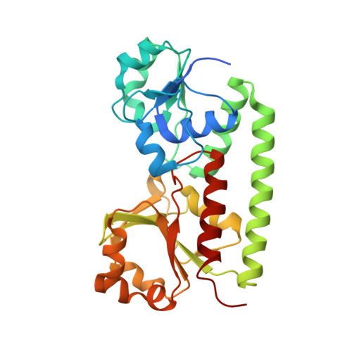 |
UniProt | |||||
Find proteins for A0A0H3AJ03 (Vibrio cholerae serotype O1 (strain ATCC 39541 / Classical Ogawa 395 / O395)) Explore A0A0H3AJ03 Go to UniProtKB: A0A0H3AJ03 | |||||
Entity Groups | |||||
| Sequence Clusters | 30% Identity50% Identity70% Identity90% Identity95% Identity100% Identity | ||||
| UniProt Group | A0A0H3AJ03 | ||||
Sequence AnnotationsExpand | |||||
Reference Sequence | |||||
| Ligands 1 Unique | |||||
|---|---|---|---|---|---|
| ID | Chains | Name / Formula / InChI Key | 2D Diagram | 3D Interactions | |
| CL Download:Ideal Coordinates CCD File | C [auth A], D [auth N] | CHLORIDE ION Cl VEXZGXHMUGYJMC-UHFFFAOYSA-M |  | ||
| Length ( Å ) | Angle ( ˚ ) |
|---|---|
| a = 57.246 | α = 90 |
| b = 71.746 | β = 90 |
| c = 125.889 | γ = 90 |
| Software Name | Purpose |
|---|---|
| PHENIX | refinement |
| PHASER | model building |
| SCALA | data scaling |
| XDS | data processing |
| Funding Organization | Location | Grant Number |
|---|---|---|
| DAE (BRNS) | India | 2013/37B/26/brns |


