5G4V | pdb_00005g4v

Association of four two-k-turn units based on Kt-7 3bG,3nC, forming a square-shaped structure


Experimental Data Snapshot

  • Method: X-RAY DIFFRACTION
  • Resolution: 2.87 Å
  • R-Value Free: 
    0.274 (Depositor), 0.273 (DCC) 
  • R-Value Work: 
    0.212 (Depositor), 0.211 (DCC) 
  • R-Value Observed: 
    0.215 (Depositor) 

Starting Model: experimental
View more details

wwPDB Validation 3D Report Full Report

Validation slider image for 5G4V

This is version 1.1 of the entry. See complete history

Literature

A Quasi-Cyclic RNA Nano-Scale Molecular Object Constructed Using Kink Turns.

Huang, L.Lilley, D.M.J.

(2016) Nanoscale 8: 15189

  • DOI: https://doi.org/10.1039/c6nr05186c
  • Primary Citation Related Structures: 
    5G4T, 5G4U, 5G4V

  • PubMed Abstract: 

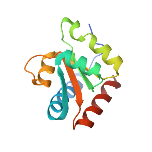
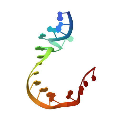
    k-Turns are widespread RNA architectural elements that mediate tertiary interactions. We describe a double-kink-turn motif comprising two inverted k-turns that forms a tight horse-shoe structure that can assemble into a variety of shapes by coaxial association of helical ends. Using X-ray crystallography we show that these assemble with two (dumbell), three (triangle) and four units (square), with or without bound protein, within the crystal lattice. In addition, exchange of a single basepair can almost double the pore radius or shape of a molecular assembly. On the basis of this analysis we synthesized a 114 nt self-complementary RNA containing six k-turns. The crystal structure of this species shows that it forms a quasi-cyclic triangular object. These are randomly disposed about the three-fold axis in the crystal lattice, generating a circular RNA of quasi D3 symmetry with a shape reminiscent of that of a cyclohexane molecule in its chair conformation. This work demonstrates that the k-turn is a powerful building block in the construction of nano-scale molecular objects, and illustrates why k-turns are widely used in natural RNA molecules to organize long-range architecture and mediate tertiary contacts.


  • Organizational Affiliation
    • Cancer Research UK Nucleic Acid Structure Research Group, MSI/WTB Complex, The University of Dundee, Dow Street, Dundee DD1 5EH, UK. d.m.j.lilley@dundee.ac.uk l.y.huang@dundee.ac.uk.

Macromolecule Content 

  • Total Structure Weight: 78.93 kDa 
  • Atom Count: 5,280 
  • Modeled Residue Count: 544 
  • Deposited Residue Count: 568 
  • Unique protein chains: 1
  • Unique nucleic acid chains: 1

Macromolecules


Find similar proteins by:|  3D Structure
Entity ID: 2
MoleculeChains  Sequence LengthOrganismDetailsImage
50S RIBOSOMAL PROTEIN L7AE
C, D, G, H
123Archaeoglobus fulgidusMutation(s): 0 
UniProt
Find proteins for O29494 (Archaeoglobus fulgidus (strain ATCC 49558 / DSM 4304 / JCM 9628 / NBRC 100126 / VC-16))
Explore O29494 
Go to UniProtKB:  O29494
Entity Groups
Sequence Clusters30% Identity50% Identity70% Identity90% Identity95% Identity100% Identity
UniProt GroupO29494
Sequence Annotations
Expand
Reference Sequence
Find similar nucleic acids by:  Sequence
Entity ID: 1
MoleculeChains LengthOrganismImage
HMKT-7
A, B, E, F
19Haloarcula marismortui
Sequence Annotations
Expand
Reference Sequence

Experimental Data & Validation

Experimental Data

  • Method: X-RAY DIFFRACTION
  • Resolution: 2.87 Å
  • R-Value Free:  0.274 (Depositor), 0.273 (DCC) 
  • R-Value Work:  0.212 (Depositor), 0.211 (DCC) 
  • R-Value Observed: 0.215 (Depositor) 
Space Group: C 1 2 1
Unit Cell:
Length ( Å )Angle ( ˚ )
a = 118.65α = 90
b = 70.64β = 105.48
c = 92.82γ = 90
Software Package:
Software NamePurpose
PHENIXrefinement
xia2data reduction
xia2data scaling

Structure Validation

View Full Validation Report


Entry History 

Deposition Data

Revision History  (Full details and data files)

  • Version 1.0: 2016-09-28
    Type: Initial release
  • Version 1.1: 2024-01-10
    Changes: Data collection, Database references, Other, Refinement description