


Crystal Structure of a Hypothetical Domain in Wnk1
Pinkas, D.M., Bufton, J.C., Sanvitale, C.E., Bartual, S.G., Adamson, R.J., Burgess-Brown, N.A., Krojer, T., von Delft, F., Arrowsmith, C.H., Edwards, A.M., Bountra, C., Bullock, A.To be published.
Experimental Data Snapshot
Starting Model: experimental
View more details
wwPDB Validation 3D Report Full Report

Entity ID: 1 | |||||
|---|---|---|---|---|---|
| Molecule | Chains | Sequence Length | Organism | Details | Image |
| WNK1 | 89 | Homo sapiens | Mutation(s): 0 | 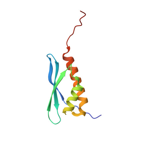 | |
| Length ( Å ) | Angle ( ˚ ) |
|---|---|
| a = 39.238 | α = 90 |
| b = 70.324 | β = 90 |
| c = 82.255 | γ = 90 |
| Software Name | Purpose |
|---|---|
| PHENIX | refinement |
| XDS | data reduction |
| XSCALE | data scaling |
| PHENIX | phasing |


