Distinct Structural Pathways Coordinate the Activation of Ampa Receptor-Auxiliary Subunit Complexes.
Dawe, G.B., Musgaard, M., Aurousseau, M.R.P., Nayeem, N., Green, T., Biggin, P.C., Bowie, D.(2016) Neuron 89: 1264
- PubMed: 26924438 Search on PubMedSearch on PubMed Central
- DOI: https://doi.org/10.1016/j.neuron.2016.01.038
- Primary Citation Related Structures:
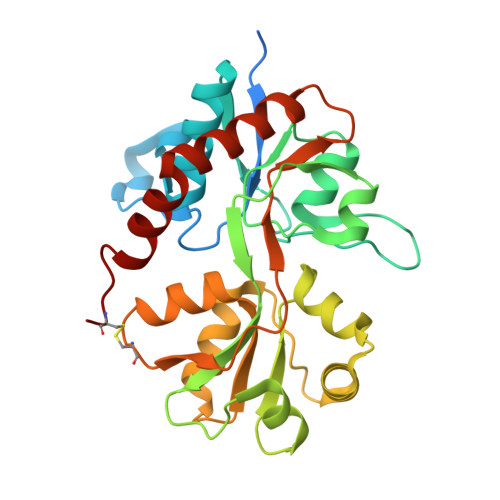
5FTH, 5FTI - PubMed Abstract:
Neurotransmitter-gated ion channels adopt different gating modes to fine-tune signaling at central synapses. At glutamatergic synapses, high and low activity of AMPA receptors (AMPARs) is observed when pore-forming subunits coassemble with or without auxiliary subunits, respectively. Whether a common structural pathway accounts for these different gating modes is unclear. Here, we identify two structural motifs that determine the time course of AMPAR channel activation. A network of electrostatic interactions at the apex of the AMPAR ligand-binding domain (LBD) is essential for gating by pore-forming subunits, whereas a conserved motif on the lower, D2 lobe of the LBD prolongs channel activity when auxiliary subunits are present. Accordingly, channel activity is almost entirely abolished by elimination of the electrostatic network but restored via auxiliary protein interactions at the D2 lobe. In summary, we propose that activation of native AMPAR complexes is coordinated by distinct structural pathways, favored by the association/dissociation of auxiliary subunits.
- Integrated Program in Neuroscience, McGill University, Montréal, QC H3A 2B4, Canada; Department of Pharmacology and Therapeutics, McGill University, Montréal, QC H3G 1Y6, Canada.
Organizational Affiliation: