5FSZ

Crystal structure of Trypanosoma cruzi macrodomain


Experimental Data Snapshot

  • Method: X-RAY DIFFRACTION
  • Resolution: 2.00 Å
  • R-Value Free: 0.258 
  • R-Value Work: 0.188 
  • R-Value Observed: 0.192 

wwPDB Validation   3D Report Full Report


This is version 1.2 of the entry. See complete history


Literature

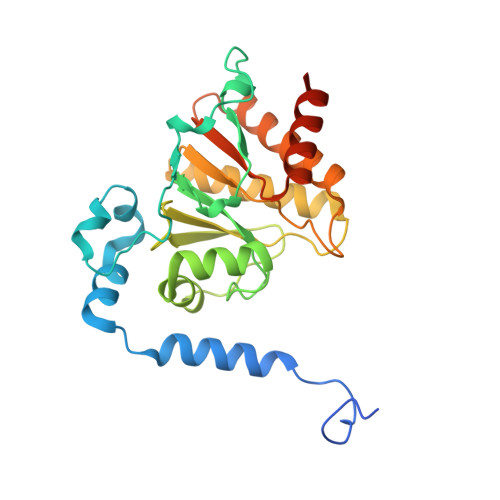
Proximal Adp-Ribose Hydrolysis in Trypanosomatids is Catalyzed by a Macrodomain.

Haikarainen, T.Lehtio, L.

(2016) Sci Rep 6: 24213

  • DOI: https://doi.org/10.1038/srep24213
  • Primary Citation of Related Structures:  
    5FSU, 5FSV, 5FSX, 5FSY, 5FSZ

  • PubMed Abstract: 

    ADP-ribosylation is a ubiquitous protein modification utilized by both prokaryotes and eukaryotes for several cellular functions, such as DNA repair, proliferation, and cell signaling. Higher eukaryotes, such as humans, utilize various enzymes to reverse the modification and to regulate ADP-ribose dependent signaling. In contrast, some lower eukaryotes, including trypanosomatids, lack many of these enzymes and therefore have a much more simplified ADP-ribose metabolism. Here we identified and characterized ADP-ribose hydrolases from Trypanosoma brucei and Trypanosoma cruzi, which are homologous to human O-acetyl-ADP-ribose deacetylases MacroD1 and MacroD2. The enzymes are capable for hydrolysis of protein linked ADP-ribose and a product of sirtuin-mediated lysine deacetylation, O-acetyl-ADP-ribose. Crystal structures of the trypanosomatid macrodomains revealed a conserved catalytic site with distinct differences to human MacroD1 and MacroD2.


  • Organizational Affiliation

    Biocenter Oulu and Faculty of Biochemistry and Molecular Medicine, University of Oulu, FI-90014 Oulu, Finland.


Macromolecules
Find similar proteins by:  (by identity cutoff)  |  3D Structure
Entity ID: 1
MoleculeChains Sequence LengthOrganismDetailsImage
MACRODOMAIN268Trypanosoma cruziMutation(s): 0 
UniProt
Find proteins for Q4DQ03 (Trypanosoma cruzi (strain CL Brener))
Explore Q4DQ03 
Go to UniProtKB:  Q4DQ03
Entity Groups  
Sequence Clusters30% Identity50% Identity70% Identity90% Identity95% Identity100% Identity
UniProt GroupQ4DQ03
Sequence Annotations
Expand
  • Reference Sequence
Experimental Data & Validation

Experimental Data

  • Method: X-RAY DIFFRACTION
  • Resolution: 2.00 Å
  • R-Value Free: 0.258 
  • R-Value Work: 0.188 
  • R-Value Observed: 0.192 
  • Space Group: P 42 21 2
Unit Cell:
Length ( Å )Angle ( ˚ )
a = 74.82α = 90
b = 74.82β = 90
c = 75.44γ = 90
Software Package:
Software NamePurpose
REFMACrefinement
XDSdata reduction
XSCALEdata scaling
PHASERphasing

Structure Validation

View Full Validation Report


Entry History 

Deposition Data

Revision History  (Full details and data files)

  • Version 1.0: 2016-04-20
    Type: Initial release
  • Version 1.1: 2017-09-13
    Changes: Data collection
  • Version 1.2: 2024-01-10
    Changes: Data collection, Database references, Other, Refinement description