5FR0

The details of glycolipid glycan hydrolysis by the structural analysis of a family 123 glycoside hydrolase from Clostridium perfringens


Experimental Data Snapshot

  • Method: X-RAY DIFFRACTION
  • Resolution: 1.75 Å
  • R-Value Free: 0.221 
  • R-Value Work: 0.181 
  • R-Value Observed: 0.183 

wwPDB Validation   3D Report Full Report


Ligand Structure Quality Assessment 


This is version 1.4 of the entry. See complete history


Literature

The Details of Glycolipid Glycan Hydrolysis by the Structural Analysis of a Family 123 Glycoside Hydrolase from Clostridium Perfringens

Noach, I.Pluvinage, B.Laurie, C.Abe, K.T.Alteen, M.Vocadlo, D.J.Boraston, A.B.

(2016) J Mol Biol 428: 3253

  • DOI: https://doi.org/10.1016/j.jmb.2016.03.020
  • Primary Citation of Related Structures:  
    5FQE, 5FQF, 5FQG, 5FQH, 5FR0

  • PubMed Abstract: 

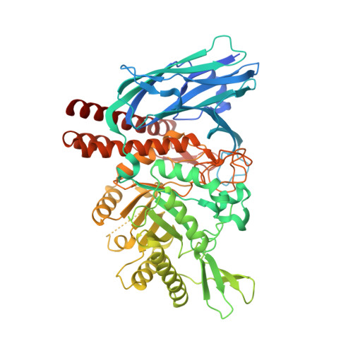
    Clostridium perfringens is an opportunistic pathogen of humans and animals whose genome encodes a wide variety of putative carbohydrate-hydrolyzing enzymes that are increasingly being shown to be directed toward the cleavage of host glycans. Among these putative enzymes is a member of glycoside hydrolase family 123. Here we show that the recombinant enzyme (referred to as CpNga123) encoded by the gene cloned from C. perfringens strain ATCC 13124 (locus tag CPF_1473) is a β-N-acetylgalactosaminidase, similar to NgaP from Paenibacillus sp. TS12. Like NgaP, CpNga123 was able to cleave the terminal β-D-GalNAc-(1→4)-D-Gal and β-D-GalNAc-(1→3)-D-Gal motifs that would be found in glycosphigolipids. The X-ray crystal structure of CpNga123 revealed it to have an N-terminal β-sandwich domain and a (β/α)8-barrel catalytic domain with a C-terminal α-helical elaboration. The structures determined in complex with reaction products provide details of the -1 subsite architecture, catalytic residues, and a structural change in the active site that is likely required to enable hydrolysis of the glycosidic bond by promoting engagement of the substrate by the catalytic residues. The features of the active site support the likelihood of a substrate-assisted catalytic mechanism for this enzyme. The structures of an inactive mutant of CpNga123 in complex with intact GA2 and Gb4 glycosphingolipid motifs reveal insight into aglycon recognition and suggest that the kinked or pleated conformation of GA2 caused by the β-1,4-linkage between N-acetylgalactosamine and galactose, and the accommodation of this conformation by the enzyme active site, may be responsible for greater activity on GA2.


  • Organizational Affiliation

    Department of Biochemistry and Microbiology, University of Victoria, Victoria, British Columbia V8W 3P6, Canada.


Macromolecules
Find similar proteins by:  (by identity cutoff)  |  3D Structure
Entity ID: 1
MoleculeChains Sequence LengthOrganismDetailsImage
BETA-N-ACETYLGALACTOSAMINIDASE610Clostridium perfringensMutation(s): 0 
UniProt
Find proteins for A0A0H2YNR7 (Clostridium perfringens (strain ATCC 13124 / DSM 756 / JCM 1290 / NCIMB 6125 / NCTC 8237 / Type A))
Explore A0A0H2YNR7 
Go to UniProtKB:  A0A0H2YNR7
Entity Groups  
Sequence Clusters30% Identity50% Identity70% Identity90% Identity95% Identity100% Identity
UniProt GroupA0A0H2YNR7
Sequence Annotations
Expand
  • Reference Sequence
Small Molecules
Ligands 2 Unique
IDChains Name / Formula / InChI Key2D Diagram3D Interactions
SIZ
Query on SIZ

Download Ideal Coordinates CCD File 
B [auth A]2-deoxy-2-[(difluoroacetyl)amino]-beta-D-galactopyranose
C8 H13 F2 N O6
QWUDJWPZSGMAGG-UXDJRKLDSA-N
PO4
Query on PO4

Download Ideal Coordinates CCD File 
C [auth A]PHOSPHATE ION
O4 P
NBIIXXVUZAFLBC-UHFFFAOYSA-K
Experimental Data & Validation

Experimental Data

  • Method: X-RAY DIFFRACTION
  • Resolution: 1.75 Å
  • R-Value Free: 0.221 
  • R-Value Work: 0.181 
  • R-Value Observed: 0.183 
  • Space Group: P 32 2 1
Unit Cell:
Length ( Å )Angle ( ˚ )
a = 99.53α = 90
b = 99.53β = 90
c = 95.31γ = 120
Software Package:
Software NamePurpose
REFMACrefinement
MOSFLMdata reduction
SCALAdata scaling
PHASERphasing

Structure Validation

View Full Validation Report


Ligand Structure Quality Assessment 


Entry History 

Deposition Data

Revision History  (Full details and data files)

  • Version 1.0: 2016-03-30
    Type: Initial release
  • Version 1.1: 2016-04-13
    Changes: Database references
  • Version 1.2: 2016-08-24
    Changes: Database references
  • Version 1.3: 2020-07-29
    Type: Remediation
    Reason: Carbohydrate remediation
    Changes: Derived calculations, Other, Structure summary
  • Version 1.4: 2024-01-10
    Changes: Data collection, Database references, Refinement description, Structure summary