Structural Basis of Lipid Targeting and Destruction by the Type V Secretion System of Pseudomonas Aeruginosa.
Da Mata Madeira, P.V., Zouhir, S., Basso, P., Neves, D., Laubier, A., Salacha, R., Bleves, S., Faudry, E., Contreras-Martel, C., Dessen, A.(2016) J Mol Biology 428: 1790
- PubMed: 27012424 Search on PubMed
- DOI: https://doi.org/10.1016/j.jmb.2016.03.012
- Primary Citation Related Structures:
5FQU, 5FYA - PubMed Abstract:
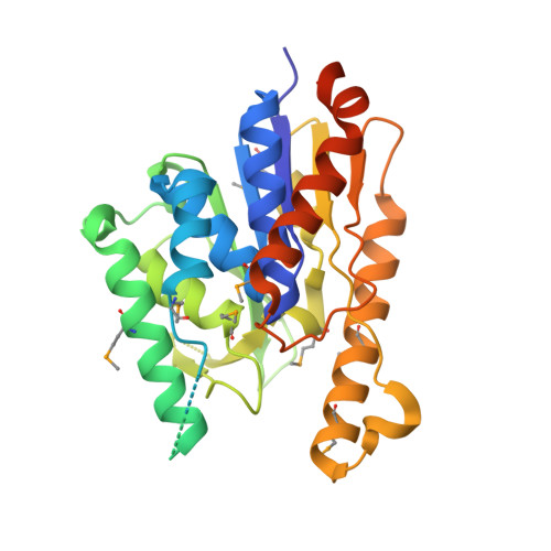
The type V secretion system is a macromolecular machine employed by a number of bacteria to secrete virulence factors into the environment. The human pathogen Pseudomonas aeruginosa employs the newly described type Vd secretion system to secrete a soluble variant of PlpD, a lipase of the patatin-like family synthesized as a single macromolecule that also carries a polypeptide transport-associated domain and a 16-stranded β-barrel. Here we report the crystal structure of the secreted form of PlpD in its biologically active state. PlpD displays a classical lipase α/β hydrolase fold with a catalytic site located within a highly hydrophobic channel that entraps a lipidic molecule. The active site is covered by a flexible lid, as in other lipases, indicating that this region in PlpD must modify its conformation in order for catalysis at the water-lipid interface to occur. PlpD displays phospholipase A1 activity and is able to recognize a number of phosphatidylinositols and other phosphatidyl analogs. PlpD is the first example of an active phospholipase secreted through the type V secretion system, for which there are more than 200 homologs, revealing details of the lipid destruction arsenal expressed by P. aeruginosa in order to establish infection.
- Brazilian National Laboratory for Biosciences (LNBio), CNPEM, Campinas, São Paulo, Brazil.
Organizational Affiliation: