Unveiling the Basis of Alkaline Stability of an Evolved Versatile Peroxidase.
Saez-Jimenez, V., Acebes, S., Garcia-Ruiz, E., Romero, A., Guallar, V., Alcalde, M., Medrano, F.J., Martinez, A.T., Ruiz-Duenas, F.J.(2016) Biochem J 473: 1917
- PubMed: 27118867 Search on PubMed
- DOI: https://doi.org/10.1042/BCJ20160248
- Primary Citation Related Structures:
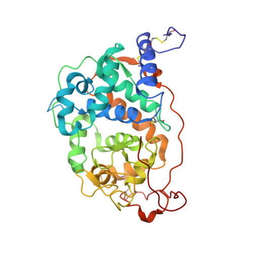
5FNB, 5FNE - PubMed Abstract:
A variant of high biotechnological interest (called 2-1B) was obtained by directed evolution of the Pleurotus eryngii VP (versatile peroxidase) expressed in Saccharomyces cerevisiae [García-Ruiz, González-Pérez, Ruiz-Dueñas, Martínez and Alcalde (2012) Biochem. J. 441: , 487-498]. 2-1B shows seven mutations in the mature protein that resulted in improved functional expression, activity and thermostability, along with a remarkable stronger alkaline stability (it retains 60% of the initial activity after 120 h of incubation at pH 9 compared with complete inactivation of the native enzyme after only 1 h). The latter is highly demanded for biorefinery applications. In the present study we investigate the structural basis behind the enhanced alkaline stabilization of this evolved enzyme. In order to do this, several VP variants containing one or several of the mutations present in 2-1B were expressed in Escherichia coli, and their alkaline stability and biochemical properties were determined. In addition, the crystal structures of 2-1B and one of the intermediate variants were solved and carefully analysed, and molecular dynamics simulations were carried out. We concluded that the introduction of three basic residues in VP (Lys-37, Arg-39 and Arg-330) led to new connections between haem and helix B (where the distal histidine residue is located), and formation of new electrostatic interactions, that avoided the hexa-co-ordination of the haem iron. These new structural determinants stabilized the haem and its environment, helping to maintain the structural enzyme integrity (with penta-co-ordinated haem iron) under alkaline conditions. Moreover, the reinforcement of the solvent-exposed area around Gln-305 in the proximal side, prompted by the Q202L mutation, further enhanced the stability.
- Centro de Investigaciones Biológicas, CSIC, Ramiro de Maeztu 9, E-28040 Madrid, Spain.
Organizational Affiliation: