5FL7 | pdb_00005fl7

Structure of the F1c10 complex from Yarrowia lipolytica ATP synthase


Experimental Data Snapshot

  • Method: X-RAY DIFFRACTION
  • Resolution: 3.50 Å
  • R-Value Free: 
    0.305 (Depositor), 0.311 (DCC) 
  • R-Value Work: 
    0.274 (Depositor), 0.278 (DCC) 
  • R-Value Observed: 
    0.275 (Depositor) 

Starting Model: experimental
View more details

wwPDB Validation 3D Report Full Report

Validation slider image for 5FL7

Ligand Structure Quality Assessment 


This is version 1.3 of the entry. See complete history

Literature

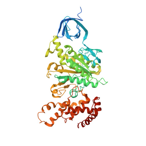
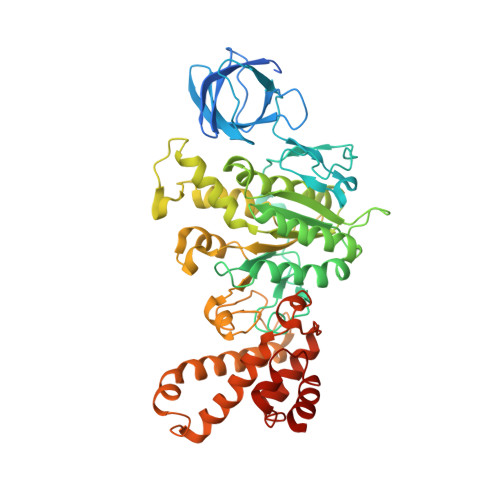
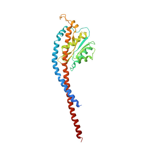
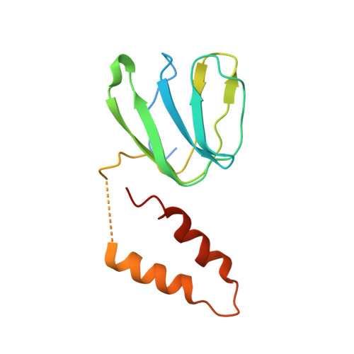
Structure of a Complete ATP Synthase Dimer Reveals the Molecular Basis of Inner Mitochondrial Membrane Morphology.

Hahn, A.Parey, K.Bublitz, M.Mills, D.J.Zickermann, V.Vonck, J.Kuhlbrandt, W.Meier, T.

(2016) Mol Cell 63: 445

  • DOI: https://doi.org/10.1016/j.molcel.2016.05.037
  • Primary Citation Related Structures: 
    5FL7

  • PubMed Abstract: 

    We determined the structure of a complete, dimeric F1Fo-ATP synthase from yeast Yarrowia lipolytica mitochondria by a combination of cryo-EM and X-ray crystallography. The final structure resolves 58 of the 60 dimer subunits. Horizontal helices of subunit a in Fo wrap around the c-ring rotor, and a total of six vertical helices assigned to subunits a, b, f, i, and 8 span the membrane. Subunit 8 (A6L in human) is an evolutionary derivative of the bacterial b subunit. On the lumenal membrane surface, subunit f establishes direct contact between the two monomers. Comparison with a cryo-EM map of the F1Fo monomer identifies subunits e and g at the lateral dimer interface. They do not form dimer contacts but enable dimer formation by inducing a strong membrane curvature of ∼100°. Our structure explains the structural basis of cristae formation in mitochondria, a landmark signature of eukaryotic cell morphology.


  • Organizational Affiliation
    • Department of Structural Biology, Max Planck Institute of Biophysics, Max-von-Laue-Str. 3, 60438 Frankfurt am Main, Germany.

Macromolecule Content 

  • Total Structure Weight: 466.47 kDa 
  • Atom Count: 30,119 
  • Modeled Residue Count: 3,996 
  • Deposited Residue Count: 4,341 
  • Unique protein chains: 6

Macromolecules

Find similar proteins by:|  3D Structure
Entity ID: 1
MoleculeChains  Sequence LengthOrganismDetailsImage
ATP SYNTHASE SUBUNIT ALPHA
A, B, C
536Yarrowia lipolyticaMutation(s): 0 
Membrane Entity: Yes 
UniProt
Find proteins for Q6C326 (Yarrowia lipolytica (strain CLIB 122 / E 150))
Explore Q6C326 
Go to UniProtKB:  Q6C326
Entity Groups
Sequence Clusters30% Identity50% Identity70% Identity90% Identity95% Identity100% Identity
UniProt GroupQ6C326
Sequence Annotations
Expand
Reference Sequence
Find similar proteins by:|  3D Structure
Entity ID: 2
MoleculeChains  Sequence LengthOrganismDetailsImage
ATP SYNTHASE SUBUNIT BETA
D, E, F
509Yarrowia lipolyticaMutation(s): 0 
EC: 3.6.3.14 (PDB Primary Data), 7.1.2.2 (UniProt)
Membrane Entity: Yes 
UniProt
Find proteins for Q6CFT7 (Yarrowia lipolytica (strain CLIB 122 / E 150))
Explore Q6CFT7 
Go to UniProtKB:  Q6CFT7
Entity Groups
Sequence Clusters30% Identity50% Identity70% Identity90% Identity95% Identity100% Identity
UniProt GroupQ6CFT7
Sequence Annotations
Expand
Reference Sequence
Find similar proteins by:|  3D Structure
Entity ID: 3
MoleculeChains  Sequence LengthOrganismDetailsImage
ATP SYNTHASE SUBUNIT GAMMA CHAIN, MITOCHONDRIAL293Yarrowia lipolyticaMutation(s): 0 
Membrane Entity: Yes 
UniProt
Find proteins for Q6C338 (Yarrowia lipolytica (strain CLIB 122 / E 150))
Explore Q6C338 
Go to UniProtKB:  Q6C338
Entity Groups
Sequence Clusters30% Identity50% Identity70% Identity90% Identity95% Identity100% Identity
UniProt GroupQ6C338
Sequence Annotations
Expand
Reference Sequence
Find similar proteins by:|  3D Structure
Entity ID: 4
MoleculeChains  Sequence LengthOrganismDetailsImage
ATP SYNTHASE DELTA CHAIN, MITOCHONDRIAL137Yarrowia lipolyticaMutation(s): 0 
Membrane Entity: Yes 
UniProt
Find proteins for Q6C877 (Yarrowia lipolytica (strain CLIB 122 / E 150))
Explore Q6C877 
Go to UniProtKB:  Q6C877
Entity Groups
Sequence Clusters30% Identity50% Identity70% Identity90% Identity95% Identity100% Identity
UniProt GroupQ6C877
Sequence Annotations
Expand
Reference Sequence
Find similar proteins by:  Sequence   |   3D Structure  
Entity ID: 5
MoleculeChains  Sequence LengthOrganismDetailsImage
ATP SYNTHASE EPSILON CHAIN, MITOCHONDRIAL16Yarrowia lipolyticaMutation(s): 0 
Find similar proteins by:|  3D Structure
Entity ID: 6
MoleculeChains  Sequence LengthOrganismDetailsImage
ATP SYNTHASE SUBUNIT 9, MITOCHONDRIAL76Yarrowia lipolyticaMutation(s): 0 
EC: 3.6.3.14
Membrane Entity: Yes 
UniProt
Find proteins for Q37695 (Yarrowia lipolytica (strain CLIB 122 / E 150))
Explore Q37695 
Go to UniProtKB:  Q37695
Entity Groups
Sequence Clusters30% Identity50% Identity70% Identity90% Identity95% Identity100% Identity
UniProt GroupQ37695
Sequence Annotations
Expand
Reference Sequence

Small Molecules

Ligands 3 Unique
IDChains Name / Formula / InChI Key2D Diagram3D Interactions
ATP

Query on ATP


Download:Ideal Coordinates CCD File
T [auth A],
V [auth B],
X [auth C]
ADENOSINE-5'-TRIPHOSPHATE
C10 H16 N5 O13 P3
ZKHQWZAMYRWXGA-KQYNXXCUSA-N
ADP

Query on ADP


Download:Ideal Coordinates CCD File
BA [auth F],
Z [auth D]
ADENOSINE-5'-DIPHOSPHATE
C10 H15 N5 O10 P2
XTWYTFMLZFPYCI-KQYNXXCUSA-N
MG

Query on MG


Download:Ideal Coordinates CCD File
AA [auth D],
CA [auth F],
U [auth A],
W [auth B],
Y [auth C]
MAGNESIUM ION
Mg
JLVVSXFLKOJNIY-UHFFFAOYSA-N

Experimental Data & Validation

Experimental Data

  • Method: X-RAY DIFFRACTION
  • Resolution: 3.50 Å
  • R-Value Free:  0.305 (Depositor), 0.311 (DCC) 
  • R-Value Work:  0.274 (Depositor), 0.278 (DCC) 
  • R-Value Observed: 0.275 (Depositor) 
Space Group: P 21 21 2
Unit Cell:
Length ( Å )Angle ( ˚ )
a = 169.5α = 90
b = 182.2β = 90
c = 193γ = 90
Software Package:
Software NamePurpose
PHENIXrefinement
XDSdata reduction
XSCALEdata scaling
PHASERphasing

Structure Validation

View Full Validation Report


Ligand Structure Quality Assessment 


Entry History 

Deposition Data

Revision History  (Full details and data files)

  • Version 1.0: 2016-06-29
    Type: Initial release
  • Version 1.1: 2016-07-20
    Changes: Database references
  • Version 1.2: 2016-08-24
    Changes: Database references
  • Version 1.3: 2024-01-10
    Changes: Data collection, Database references, Derived calculations, Other, Refinement description