5FIR | pdb_00005fir

Crystal structure of C. elegans XRN2 in complex with the XRN2-binding domain of PAXT-1


Experimental Data Snapshot

  • Method: X-RAY DIFFRACTION
  • Resolution: 2.84 Å
  • R-Value Free: 
    0.238 (Depositor), 0.244 (DCC) 
  • R-Value Work: 
    0.185 (Depositor), 0.196 (DCC) 
  • R-Value Observed: 
    0.187 (Depositor) 

Starting Model: in silico
View more details

wwPDB Validation 3D Report Full Report

Validation slider image for 5FIR

This is version 1.4 of the entry. See complete history

Literature

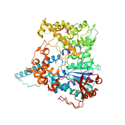
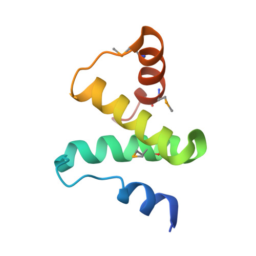
Structural Basis and Function of Xrn2-Binding by Xtb Domains

Richter, H.Katic, I.Gut, H.Grosshans, H.

(2016) Nat Struct Mol Biol 23: 164

  • DOI: https://doi.org/10.1038/nsmb.3155
  • Primary Citation Related Structures: 
    5FIR

  • PubMed Abstract: 

    The RNase XRN2 is essential in RNA metabolism. In Caenorhabditis elegans, XRN2 functions with PAXT-1, which shares a putative XRN2-binding domain (XTBD) with otherwise unrelated mammalian proteins. Here, we characterize the structure and function of an XTBD-XRN2 complex. Although XTBD stably interconnects two XRN2 domains through numerous interacting residues, mutation of a single critical residue suffices to disrupt XTBD-XRN2 complexes in vitro and to recapitulate paxt-1-null mutant phenotypes in vivo. Demonstrating conservation of function, vertebrate XTBD-containing proteins bind XRN2 in vitro, and human CDKN2AIPNL (HsC2AIL) can substitute for PAXT-1 in vivo. In vertebrates, which express three distinct XTBD-containing proteins, XRN2 may partition into distinct stable heterodimeric complexes, which probably differ in subcellular localization or function. In C. elegans, complex formation with PAXT-1, the sole XTBD protein, serves to preserve the stability of XRN2 in the absence of substrate.


  • Organizational Affiliation
    • Friedrich Miescher Institute for Biomedical Research, Basel, Switzerland.

Macromolecule Content 

  • Total Structure Weight: 505.98 kDa 
  • Atom Count: 33,550 
  • Modeled Residue Count: 4,073 
  • Deposited Residue Count: 4,284 
  • Unique protein chains: 2

Macromolecules

Find similar proteins by:|  3D Structure
Entity ID: 1
MoleculeChains  Sequence LengthOrganismDetailsImage
5'-3' EXORIBONUCLEASE 2 HOMOLOG
A, C, E, G, I
A, C, E, G, I, K
636Caenorhabditis elegansMutation(s): 0 
Gene Names: XRN-2Y48B6A.3
EC: 3.1.13
UniProt
Find proteins for Q9U299 (Caenorhabditis elegans)
Explore Q9U299 
Go to UniProtKB:  Q9U299
Entity Groups
Sequence Clusters30% Identity50% Identity70% Identity90% Identity95% Identity100% Identity
UniProt GroupQ9U299
Sequence Annotations
Expand
Reference Sequence
Find similar proteins by:|  3D Structure
Entity ID: 2
MoleculeChains  Sequence LengthOrganismDetailsImage
PAXT-1
B, D, F, H, J
B, D, F, H, J, L
78Caenorhabditis elegansMutation(s): 0 
Gene Names: PAXT-1CELE_R05D11.6R05D11.6
UniProt
Find proteins for Q21738 (Caenorhabditis elegans)
Explore Q21738 
Go to UniProtKB:  Q21738
Entity Groups
Sequence Clusters30% Identity50% Identity70% Identity90% Identity95% Identity100% Identity
UniProt GroupQ21738
Sequence Annotations
Expand
Reference Sequence

Small Molecules

Ligands 1 Unique
IDChains Name / Formula / InChI Key2D Diagram3D Interactions
SO4

Query on SO4


Download:Ideal Coordinates CCD File
AA [auth E]
BA [auth E]
CA [auth G]
DA [auth G]
EA [auth G]
AA [auth E],
BA [auth E],
CA [auth G],
DA [auth G],
EA [auth G],
FA [auth G],
GA [auth I],
HA [auth I],
IA [auth I],
JA [auth K],
KA [auth K],
LA [auth K],
M [auth A],
MA [auth K],
N [auth A],
O [auth A],
P [auth A],
Q [auth A],
R [auth A],
S [auth A],
T [auth A],
U [auth C],
V [auth C],
W [auth C],
X [auth E],
Y [auth E],
Z [auth E]
SULFATE ION
O4 S
QAOWNCQODCNURD-UHFFFAOYSA-L
Modified Residues  1 Unique
IDChains TypeFormula2D DiagramParent
MSE
Query on MSE
A, C, E, G, I
A, C, E, G, I, K
L-PEPTIDE LINKINGC5 H11 N O2 SeMET

Experimental Data & Validation

Experimental Data

  • Method: X-RAY DIFFRACTION
  • Resolution: 2.84 Å
  • R-Value Free:  0.238 (Depositor), 0.244 (DCC) 
  • R-Value Work:  0.185 (Depositor), 0.196 (DCC) 
  • R-Value Observed: 0.187 (Depositor) 
Space Group: P 21 21 21
Unit Cell:
Length ( Å )Angle ( ˚ )
a = 170.21α = 90
b = 200.77β = 90
c = 202.99γ = 90
Software Package:
Software NamePurpose
PHENIXrefinement
XDSdata reduction
XSCALEdata scaling
PHASERphasing

Structure Validation

View Full Validation Report


Entry History 

Deposition Data

Revision History  (Full details and data files)

  • Version 1.0: 2016-01-20
    Type: Initial release
  • Version 1.1: 2016-01-27
    Changes: Database references
  • Version 1.2: 2016-03-02
    Changes: Database references
  • Version 1.3: 2024-05-01
    Changes: Data collection, Database references, Derived calculations, Other, Refinement description
  • Version 1.4: 2024-11-20
    Changes: Structure summary