5FHH | pdb_00005fhh

Structure of human Pif1 helicase domain residues 200-641


Experimental Data Snapshot

  • Method: X-RAY DIFFRACTION
  • Resolution: 3.60 Å
  • R-Value Free: 
    0.355 (Depositor), 0.361 (DCC) 
  • R-Value Work: 
    0.310 (Depositor), 0.323 (DCC) 
  • R-Value Observed: 
    0.313 (Depositor) 

wwPDB Validation 3D Report Full Report

Validation slider image for 5FHH

Ligand Structure Quality Assessment 


This is version 1.1 of the entry. See complete history

Literature

Structural and Functional Insights into the Unwinding Mechanism of Bacteroides sp Pif1

Zhou, X.Ren, W.Bharath, S.R.Tang, X.He, Y.Chen, C.Liu, Z.Li, D.Song, H.

(2016) Cell Rep 14: 2030-2039

  • DOI: https://doi.org/10.1016/j.celrep.2016.02.008
  • Primary Citation Related Structures: 
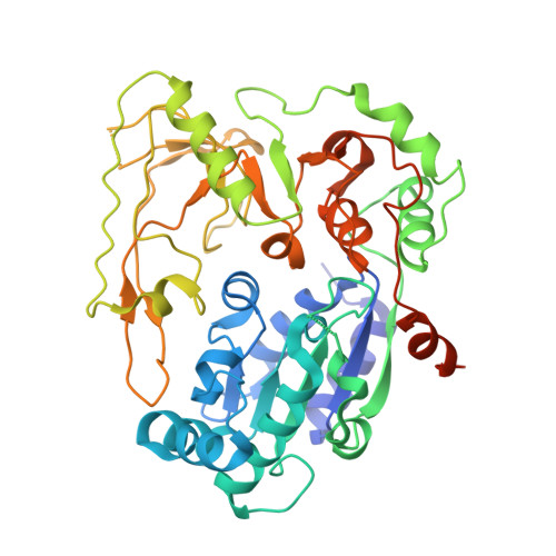
    5FHD, 5FHE, 5FHF, 5FHG, 5FHH

  • PubMed Abstract: 

    Pif1 is a conserved SF1B DNA helicase involved in maintaining genome stability through unwinding double-stranded DNAs (dsDNAs), DNA/RNA hybrids, and G quadruplex (G4) structures. Here, we report the structures of the helicase domain of human Pif1 and Bacteroides sp Pif1 (BaPif1) in complex with ADP-AlF4(-) and two different single-stranded DNAs (ssDNAs). The wedge region equivalent to the β hairpin in other SF1B DNA helicases folds into an extended loop followed by an α helix. The Pif1 signature motif of BaPif1 interacts with the wedge region and a short helix in order to stabilize these ssDNA binding elements, therefore indirectly exerting its functional role. Domain 2B of BaPif1 undergoes a large conformational change upon concomitant binding of ATP and ssDNA, which is critical for Pif1's activities. BaPif1 cocrystallized with a tailed dsDNA and ADP-AlF4(-), resulting in a bound ssDNA bent nearly 90° at the ssDNA/dsDNA junction. The conformational snapshots of BaPif1 provide insights into the mechanism governing the helicase activity of Pif1.


  • Organizational Affiliation
    • Life Sciences Institute and Innovation Center for Cell Signaling Network, Zhejiang University, 388 Yuhangtang Road, Hangzhou 310058, China; Institute of Molecular and Cell Biology, 61 Biopolis Drive, Singapore 138673, Singapore.

Macromolecule Content 

  • Total Structure Weight: 97.07 kDa 
  • Atom Count: 5,128 
  • Modeled Residue Count: 788 
  • Deposited Residue Count: 884 
  • Unique protein chains: 1

Macromolecules

Find similar proteins by:|  3D Structure
Entity ID: 1
MoleculeChains  Sequence LengthOrganismDetailsImage
ATP-dependent DNA helicase PIF1
A, B
442Homo sapiensMutation(s): 0 
Gene Names: PIF1C15orf20
EC: 3.6.4.12 (PDB Primary Data), 5.6.2.3 (UniProt)
UniProt & NIH Common Fund Data Resources
Find proteins for Q9H611 (Homo sapiens)
Explore Q9H611 
Go to UniProtKB:  Q9H611
PHAROS:  Q9H611
GTEx:  ENSG00000140451 
Entity Groups
Sequence Clusters30% Identity50% Identity70% Identity90% Identity95% Identity100% Identity
UniProt GroupQ9H611
Sequence Annotations
Expand
Reference Sequence

Experimental Data & Validation

Experimental Data

  • Method: X-RAY DIFFRACTION
  • Resolution: 3.60 Å
  • R-Value Free:  0.355 (Depositor), 0.361 (DCC) 
  • R-Value Work:  0.310 (Depositor), 0.323 (DCC) 
  • R-Value Observed: 0.313 (Depositor) 
Space Group: P 32 2 1
Unit Cell:
Length ( Å )Angle ( ˚ )
a = 204.75α = 90
b = 204.75β = 90
c = 77.791γ = 120
Software Package:
Software NamePurpose
PHENIXrefinement
Aimlessdata reduction
Aimlessdata scaling
PHASERphasing

Structure Validation

View Full Validation Report


Ligand Structure Quality Assessment 


Entry History 

Deposition Data

Revision History  (Full details and data files)

  • Version 1.0: 2016-03-30
    Type: Initial release
  • Version 1.1: 2024-11-20
    Changes: Data collection, Database references, Derived calculations, Structure summary