5FG3

Crystal structure of GDP-bound aIF5B from Aeropyrum pernix


Experimental Data Snapshot

  • Method: X-RAY DIFFRACTION
  • Resolution: 1.90 Å
  • R-Value Free: 0.243 
  • R-Value Work: 0.194 
  • R-Value Observed: 0.197 

wwPDB Validation   3D Report Full Report


Ligand Structure Quality Assessment 


This is version 1.2 of the entry. See complete history


Literature

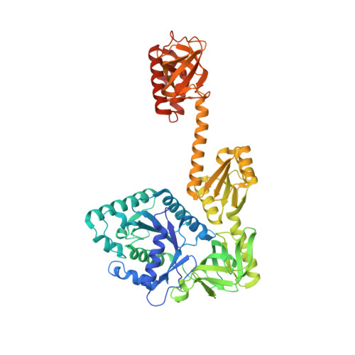
Crystal structure of translation initiation factor 5B from the crenarchaeon Aeropyrum pernix.

Murakami, R.Miyoshi, T.Uchiumi, T.Ito, K.

(2016) Proteins 84: 712-717

  • DOI: https://doi.org/10.1002/prot.25009
  • Primary Citation of Related Structures:  
    5FG3

  • PubMed Abstract: 

    Initiation factor 5B (IF5B) is a universally conserved translational GTPase that catalyzes ribosomal subunit joining. In eukaryotes, IF5B directly interacts via a groove in its domain IV with initiation factor 1A (IF1A), another universally conserved initiation factor, to accomplish efficient subunit joining. Here, we have determined the first structure of a crenarchaeal IF5B, which revealed that the archaea-specific region of IF5B (helix α15) binds and occludes the groove of domain IV. Therefore, archaeal IF5B cannot access IF1A in the same manner as eukaryotic IF5B. This fact suggests that different relationships between IF5B and IF1A exist in archaea and eukaryotes.


  • Organizational Affiliation

    Department of Food and Life Sciences, Graduate School of Science and Technology, Niigata University, Japan.


Macromolecules
Find similar proteins by:  (by identity cutoff)  |  3D Structure
Entity ID: 1
MoleculeChains Sequence LengthOrganismDetailsImage
Probable translation initiation factor IF-2623Aeropyrum pernix K1Mutation(s): 0 
Gene Names: infBAPE_2374
UniProt
Find proteins for Q9Y9B3 (Aeropyrum pernix (strain ATCC 700893 / DSM 11879 / JCM 9820 / NBRC 100138 / K1))
Explore Q9Y9B3 
Go to UniProtKB:  Q9Y9B3
Entity Groups  
Sequence Clusters30% Identity50% Identity70% Identity90% Identity95% Identity100% Identity
UniProt GroupQ9Y9B3
Sequence Annotations
Expand
  • Reference Sequence
Small Molecules
Ligands 1 Unique
IDChains Name / Formula / InChI Key2D Diagram3D Interactions
GDP
Query on GDP

Download Ideal Coordinates CCD File 
B [auth A]GUANOSINE-5'-DIPHOSPHATE
C10 H15 N5 O11 P2
QGWNDRXFNXRZMB-UUOKFMHZSA-N
Experimental Data & Validation

Experimental Data

  • Method: X-RAY DIFFRACTION
  • Resolution: 1.90 Å
  • R-Value Free: 0.243 
  • R-Value Work: 0.194 
  • R-Value Observed: 0.197 
  • Space Group: P 1 21 1
Unit Cell:
Length ( Å )Angle ( ˚ )
a = 57.853α = 90
b = 97.448β = 94.77
c = 61.296γ = 90
Software Package:
Software NamePurpose
REFMACrefinement
XDSdata collection
MOLREPphasing
XDSdata reduction
XSCALEdata scaling
Cootmodel building

Structure Validation

View Full Validation Report


Ligand Structure Quality Assessment 


Entry History & Funding Information

Deposition Data


Funding OrganizationLocationGrant Number
Japan Society for The Promotion of ScienceJapan15J04972
Japan Society for The Promotion of ScienceJapan21770108
Uchida Energy Science Promotion FoundationJapan22-1-10
The Ministry of Education,Culture,Sports,Science and TechnologyJapan24370073

Revision History  (Full details and data files)

  • Version 1.0: 2016-05-04
    Type: Initial release
  • Version 1.1: 2020-02-19
    Changes: Data collection, Database references, Derived calculations
  • Version 1.2: 2023-11-08
    Changes: Data collection, Database references, Refinement description