Structural insights into the IgE mediated responses induced by the allergens Hev b 8 and Zea m 12 in their dimeric forms.
Mares-Mejia, I., Martinez-Caballero, S., Garay-Canales, C., Cano-Sanchez, P., Torres-Larios, A., Lara-Gonzalez, S., Ortega, E., Rodriguez-Romero, A.(2016) Sci Rep 6: 32552-32552
- PubMed: 27586352 Search on PubMedSearch on PubMed Central
- DOI: https://doi.org/10.1038/srep32552
- Primary Citation Related Structures:
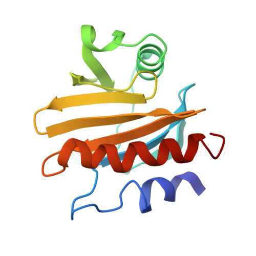
5FDS, 5FEF, 5FEG - PubMed Abstract:
Oligomerization of allergens plays an important role in IgE-mediated reactions, as effective crosslinking of IgE- FcεRI complexes on the cell membrane is dependent on the number of exposed B-cell epitopes in a single allergen molecule or on the occurrence of identical epitopes in a symmetrical arrangement. Few studies have attempted to experimentally demonstrate the connection between allergen dimerization and the ability to trigger allergic reactions. Here we studied plant allergenic profilins rHev b 8 (rubber tree) and rZea m 12 (maize) because they represent an important example of cross-reactivity in the latex-pollen-food syndrome. Both allergens in their monomeric and dimeric states were isolated and characterized by exclusion chromatography and mass spectrometry and were used in immunological in vitro experiments. Their crystal structures were solved, and for Hev b 8 a disulfide-linked homodimer was found. Comparing the structures we established that the longest loop is relevant for recognition by IgE antibodies, whereas the conserved regions are important for cross-reactivity. We produced a novel monoclonal murine IgE (mAb 2F5), specific for rHev b 8, which was useful to provide evidence that profilin dimerization considerably increases the IgE-mediated degranulation in rat basophilic leukemia cells.
- Instituto de Química, Universidad Nacional Autónoma de México, Circuito Exterior, Cd. Universitaria, Coyoacán, Ciudad de México, 04510.
Organizational Affiliation: