5F6C | pdb_00005f6c

The structure of E. coli RNase E catalytically inactive mutant with RNA bound


Experimental Data Snapshot

  • Method: X-RAY DIFFRACTION
  • Resolution: 3.00 Å
  • R-Value Free: 
    0.250 (Depositor), 0.248 (DCC) 
  • R-Value Work: 
    0.175 (Depositor), 0.178 (DCC) 
  • R-Value Observed: 
    0.179 (Depositor) 

wwPDB Validation 3D Report Full Report

Validation slider image for 5F6C

This is version 1.3 of the entry. See complete history

Literature

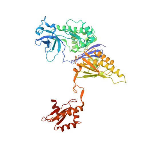
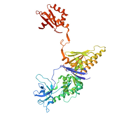
Substrate Recognition and Autoinhibition in the Central Ribonuclease RNase E.

Bandyra, K.J.Wandzik, J.M.Luisi, B.F.

(2018) Mol Cell 72: 275-285.e4

  • DOI: https://doi.org/10.1016/j.molcel.2018.08.039
  • Primary Citation Related Structures: 
    5F6C, 6G63

  • PubMed Abstract: 

    The endoribonuclease RNase E is a principal factor in RNA turnover and processing that helps to exercise fine control of gene expression in bacteria. While its catalytic activity can be strongly influenced by the chemical identity of the 5' end of RNA substrates, the enzyme can also cleave numerous substrates irrespective of the chemistry of their 5' ends through a mechanism that has remained largely unexplained. We report structural and functional data illuminating details of both operational modes. Our crystal structure of RNase E in complex with the sRNA RprA reveals a duplex recognition site that saddles an inter-protomer surface to help present substrates for cleavage. Our data also reveal an autoinhibitory pocket that modulates the overall activity of the ribonuclease. Taking these findings together, we propose how RNase E uses versatile modes of RNA recognition to achieve optimal activity and specificity.


  • Organizational Affiliation
    • Department of Biochemistry, University of Cambridge, Tennis Court Road, Cambridge CB2 1GA, UK. Electronic address: kjb69@cam.ac.uk.

Macromolecule Content 

  • Total Structure Weight: 117.32 kDa 
  • Atom Count: 8,088 
  • Modeled Residue Count: 1,027 
  • Deposited Residue Count: 1,030 
  • Unique protein chains: 2
  • Unique nucleic acid chains: 2

Macromolecules


Find similar proteins by:|  3D Structure
Entity ID: 1
MoleculeChains  Sequence LengthOrganismDetailsImage
Ribonuclease E512Escherichia coli K-12Mutation(s): 2 
Gene Names: rneamshmp1b1084JW1071
EC: 3.1.26.12
UniProt
Find proteins for P21513 (Escherichia coli (strain K12))
Explore P21513 
Go to UniProtKB:  P21513
Entity Groups
Sequence Clusters30% Identity50% Identity70% Identity90% Identity95% Identity100% Identity
UniProt GroupP21513
Sequence Annotations
Expand
Reference Sequence
Find similar proteins by:|  3D Structure
Entity ID: 2
MoleculeChains  Sequence LengthOrganismDetailsImage
Ribonuclease E513Escherichia coli K-12Mutation(s): 2 
Gene Names: rneamshmp1b1084JW1071
EC: 3.1.26.12
UniProt
Find proteins for P21513 (Escherichia coli (strain K12))
Explore P21513 
Go to UniProtKB:  P21513
Entity Groups
Sequence Clusters30% Identity50% Identity70% Identity90% Identity95% Identity100% Identity
UniProt GroupP21513
Sequence Annotations
Expand
Reference Sequence
Find similar nucleic acids by:  Sequence
Entity ID: 3
MoleculeChains LengthOrganismImage
RNA (5'-R(P*GP*U)-3')2Escherichia coli
Sequence Annotations
Expand
Reference Sequence
Find similar nucleic acids by:  Sequence
Entity ID: 4
MoleculeChains LengthOrganismImage
RNA (5'-R(P*GP*UP*G)-3')D [auth E]3Escherichia coli
Sequence Annotations
Expand
Reference Sequence

Experimental Data & Validation

Experimental Data

  • Method: X-RAY DIFFRACTION
  • Resolution: 3.00 Å
  • R-Value Free:  0.250 (Depositor), 0.248 (DCC) 
  • R-Value Work:  0.175 (Depositor), 0.178 (DCC) 
  • R-Value Observed: 0.179 (Depositor) 
Space Group: C 1 2 1
Unit Cell:
Length ( Å )Angle ( ˚ )
a = 91.619α = 90
b = 122.562β = 99.77
c = 122.195γ = 90
Software Package:
Software NamePurpose
PHENIXrefinement
MOSFLMdata reduction
Aimlessdata scaling
PHASERphasing

Structure Validation

View Full Validation Report


Entry History 

& Funding Information

Deposition Data


Funding OrganizationLocationGrant Number
Wellcome TrustUnited Kingdom076846/Z/05/A

Revision History  (Full details and data files)

  • Version 1.0: 2016-12-14
    Type: Initial release
  • Version 1.1: 2018-10-10
    Changes: Data collection, Database references, Structure summary
  • Version 1.2: 2019-02-27
    Changes: Data collection, Database references, Structure summary
  • Version 1.3: 2024-05-08
    Changes: Data collection, Database references, Derived calculations