5F5K

E.Coli GlpG Y205F mutant complexed with aldehyde inhibitor in DMPC/CHAPSO bicelle


Experimental Data Snapshot

  • Method: X-RAY DIFFRACTION
  • Resolution: 2.40 Å
  • R-Value Free: 0.288 
  • R-Value Work: 0.234 
  • R-Value Observed: 0.237 

wwPDB Validation   3D Report Full Report


This is version 1.1 of the entry. See complete history


Literature

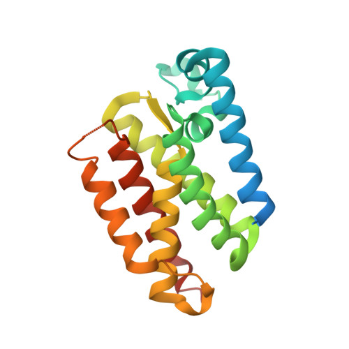
Crystal Structures and Inhibition Kinetics Reveal a Two-Stage Catalytic Mechanism with Drug Design Implications for Rhomboid Proteolysis.

Cho, S.Dickey, S.W.Urban, S.

(2016) Mol Cell 61: 329-340

  • DOI: https://doi.org/10.1016/j.molcel.2015.12.022
  • Primary Citation of Related Structures:  
    5F5B, 5F5D, 5F5G, 5F5J, 5F5K

  • PubMed Abstract: 

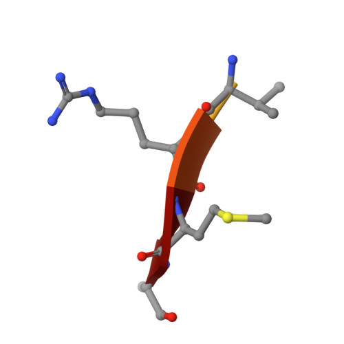
    Intramembrane proteases signal by releasing proteins from the membrane, but despite their importance, their enzymatic mechanisms remain obscure. We probed rhomboid proteases with reversible, mechanism-based inhibitors that allow precise kinetic analysis and faithfully mimic the transition state structurally. Unexpectedly, inhibition by peptide aldehydes is non-competitive, revealing that in the Michaelis complex, substrate does not contact the catalytic center. Structural analysis in a membrane revealed that all extracellular loops of rhomboid make stabilizing interactions with substrate, but mainly through backbone interactions, explaining rhomboid's broad sequence selectivity. At the catalytic site, the tetrahedral intermediate lies covalently attached to the catalytic serine alone, with the oxyanion stabilized by unusual tripartite interactions with the side chains of H150, N154, and the backbone of S201. We also visualized unexpected substrate-enzyme interactions at the non-essential P2/P3 residues. These "extra" interactions foster potent rhomboid inhibition in living cells, thereby opening avenues for rational design of selective rhomboid inhibitors.


  • Organizational Affiliation

    Howard Hughes Medical Institute, Department of Molecular Biology & Genetics, Johns Hopkins University School of Medicine, Room 507 PCTB, 725 North Wolfe Street, Baltimore, Maryland, USA, 21205.


Macromolecules
Find similar proteins by:  (by identity cutoff)  |  3D Structure
Entity ID: 1
MoleculeChains Sequence LengthOrganismDetailsImage
Rhomboid protease GlpG211Escherichia coliMutation(s): 1 
Gene Names: glpGSK83_00858
EC: 3.4.21.105
Membrane Entity: Yes 
UniProt
Find proteins for P09391 (Escherichia coli (strain K12))
Explore P09391 
Go to UniProtKB:  P09391
Entity Groups  
Sequence Clusters30% Identity50% Identity70% Identity90% Identity95% Identity100% Identity
UniProt GroupP09391
Sequence Annotations
Expand
  • Reference Sequence

Find similar proteins by:  Sequence   |   3D Structure  

Entity ID: 2
MoleculeChains Sequence LengthOrganismDetailsImage
Peptidic derivative of Gurken: ACE-ARG-LYS-VAL-ARG-MET-ALA-aldehyde7Drosophila melanogasterMutation(s): 0 
Membrane Entity: Yes 
UniProt
Find proteins for P42287 (Drosophila melanogaster)
Explore P42287 
Go to UniProtKB:  P42287
Entity Groups  
UniProt GroupP42287
Sequence Annotations
Expand
  • Reference Sequence
Small Molecules
Modified Residues  1 Unique
IDChains TypeFormula2D DiagramParent
5XU
Query on 5XU
B
L-PEPTIDE LINKINGC3 H7 N OALA
Experimental Data & Validation

Experimental Data

  • Method: X-RAY DIFFRACTION
  • Resolution: 2.40 Å
  • R-Value Free: 0.288 
  • R-Value Work: 0.234 
  • R-Value Observed: 0.237 
  • Space Group: C 2 2 21
Unit Cell:
Length ( Å )Angle ( ˚ )
a = 70.32α = 90
b = 96.12β = 90
c = 62.67γ = 90
Software Package:
Software NamePurpose
REFMACrefinement
iMOSFLMdata reduction
Aimlessdata scaling
MOLREPphasing

Structure Validation

View Full Validation Report


Entry History 

Deposition Data

Revision History  (Full details and data files)

  • Version 1.0: 2016-02-10
    Type: Initial release
  • Version 1.1: 2016-02-17
    Changes: Database references