5F4Z | pdb_00005f4z

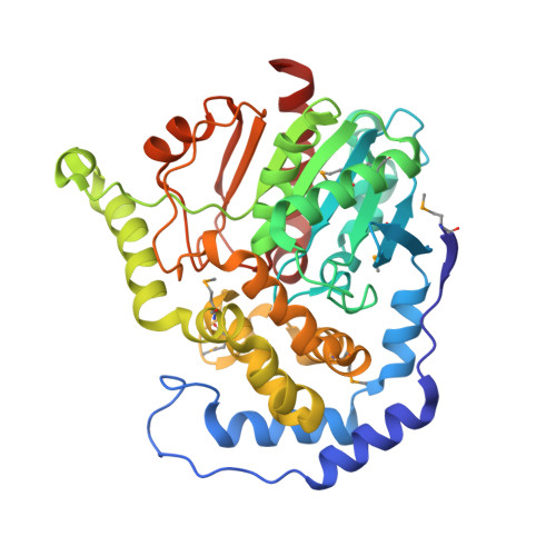
The crystal structure of an epoxide hydrolase from Streptomyces carzinostaticus subsp. neocarzinostaticus


Experimental Data Snapshot

  • Method: X-RAY DIFFRACTION
  • Resolution: 1.82 Å
  • R-Value Free: 
    0.200 (Depositor), 0.200 (DCC) 
  • R-Value Work: 
    0.160 (Depositor), 0.162 (DCC) 
  • R-Value Observed: 
    0.162 (Depositor) 

wwPDB Validation 3D Report Full Report

Validation slider image for 5F4Z

Ligand Structure Quality Assessment 


This is version 1.5 of the entry. See complete history

Literature

The crystal structure of an epoxide hydrolase from Streptomyces carzinostaticus subsp. neocarzinostaticus

Tan, K.Li, H.Jedrzejczak, R.BABNIGG, G.BINGMAN, C.A.YENNAMALLI, R.LOHMAN, J.Chang, C.Y.Shen, B.Phillips Jr., G.N.Joachimiak, A.

To be published.

Macromolecule Content 

  • Total Structure Weight: 220.82 kDa 
  • Atom Count: 16,650 
  • Modeled Residue Count: 1,944 
  • Deposited Residue Count: 1,965 
  • Unique protein chains: 1

Macromolecules

Find similar proteins by:|  3D Structure
Entity ID: 1
MoleculeChains  Sequence LengthOrganismDetailsImage
Epoxide hydrolase
A, B, C, D, E
393Streptomyces carzinostaticus subsp. neocarzinostaticusMutation(s): 0 
UniProt
Find proteins for Q84HB1 (Streptomyces carzinostaticus subsp. neocarzinostaticus)
Explore Q84HB1 
Go to UniProtKB:  Q84HB1
Entity Groups
Sequence Clusters30% Identity50% Identity70% Identity90% Identity95% Identity100% Identity
UniProt GroupQ84HB1
Sequence Annotations
Expand
Reference Sequence

Small Molecules

Ligands 6 Unique
IDChains Name / Formula / InChI Key2D Diagram3D Interactions
PG4

Query on PG4


Download:Ideal Coordinates CCD File
O [auth B]TETRAETHYLENE GLYCOL
C8 H18 O5
UWHCKJMYHZGTIT-UHFFFAOYSA-N
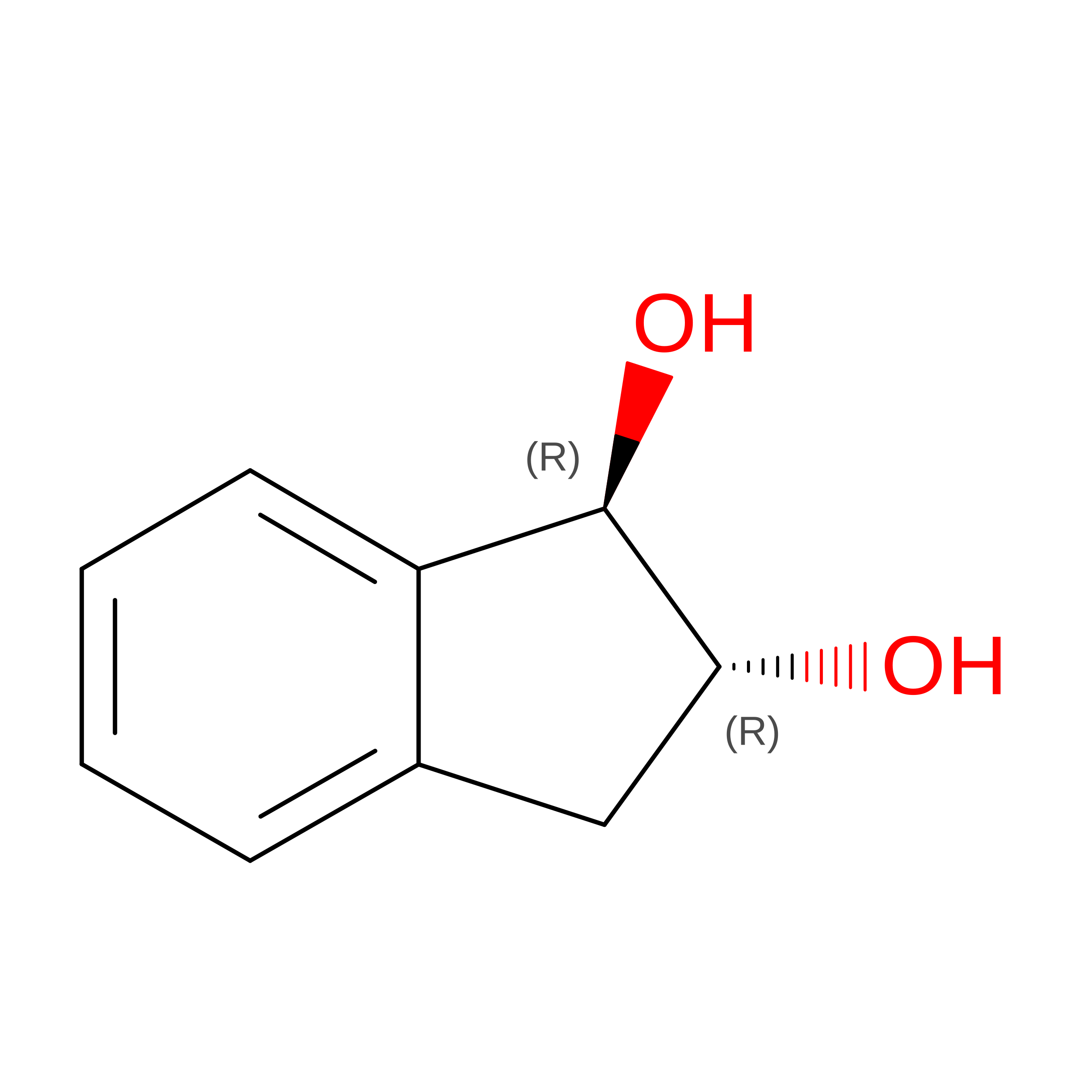
5V4

Query on 5V4


Download:Ideal Coordinates CCD File
M [auth A],
Q [auth B],
S [auth C],
T [auth D],
V [auth E]
(1~{R},2~{R})-2,3-dihydro-1~{H}-indene-1,2-diol
C9 H10 O2
YKXXBEOXRPZVCC-RKDXNWHRSA-N
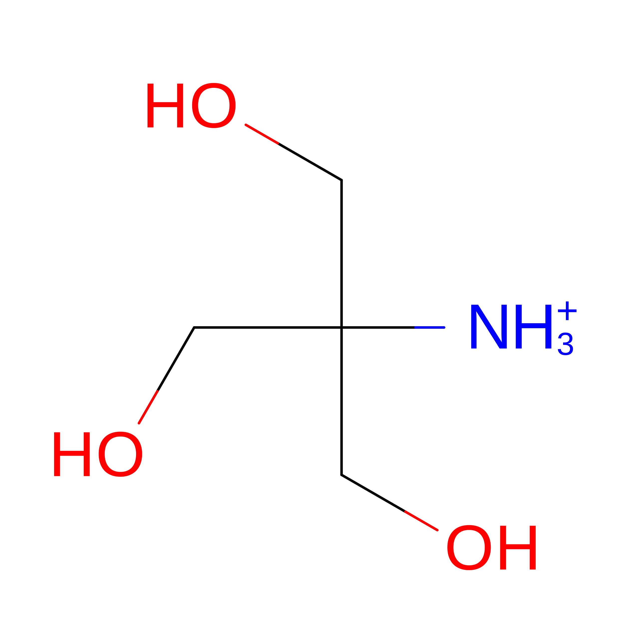
TRS

Query on TRS


Download:Ideal Coordinates CCD File
P [auth B]2-AMINO-2-HYDROXYMETHYL-PROPANE-1,3-DIOL
C4 H12 N O3
LENZDBCJOHFCAS-UHFFFAOYSA-O
PEG

Query on PEG


Download:Ideal Coordinates CCD File
L [auth A]DI(HYDROXYETHYL)ETHER
C4 H10 O3
MTHSVFCYNBDYFN-UHFFFAOYSA-N
GOL

Query on GOL


Download:Ideal Coordinates CCD File
F [auth A]
G [auth A]
H [auth A]
I [auth A]
J [auth A]
F [auth A],
G [auth A],
H [auth A],
I [auth A],
J [auth A],
R [auth C],
U [auth E]
GLYCEROL
C3 H8 O3
PEDCQBHIVMGVHV-UHFFFAOYSA-N
ACT

Query on ACT


Download:Ideal Coordinates CCD File
K [auth A],
N [auth B]
ACETATE ION
C2 H3 O2
QTBSBXVTEAMEQO-UHFFFAOYSA-M
Modified Residues  1 Unique
IDChains TypeFormula2D DiagramParent
MSE
Query on MSE
A, B, C, D, E
L-PEPTIDE LINKINGC5 H11 N O2 SeMET

Experimental Data & Validation

Experimental Data

  • Method: X-RAY DIFFRACTION
  • Resolution: 1.82 Å
  • R-Value Free:  0.200 (Depositor), 0.200 (DCC) 
  • R-Value Work:  0.160 (Depositor), 0.162 (DCC) 
  • R-Value Observed: 0.162 (Depositor) 
Space Group: C 1 2 1
Unit Cell:
Length ( Å )Angle ( ˚ )
a = 179.356α = 90
b = 93.068β = 115.33
c = 134.059γ = 90
Software Package:
Software NamePurpose
PHENIXrefinement
HKL-3000data reduction
HKL-3000data scaling

Structure Validation

View Full Validation Report


Ligand Structure Quality Assessment 


Entry History 

& Funding Information

Deposition Data


Funding OrganizationLocationGrant Number
National Institutes of Health/National Institute of General Medical Sciences (NIH/NIGMS)United StatesGM115586

Revision History  (Full details and data files)

  • Version 1.0: 2016-02-17
    Type: Initial release
  • Version 1.1: 2017-09-27
    Changes: Author supporting evidence, Derived calculations
  • Version 1.2: 2019-02-06
    Changes: Data collection, Source and taxonomy
  • Version 1.3: 2019-12-25
    Changes: Author supporting evidence
  • Version 1.4: 2020-09-23
    Changes: Database references, Structure summary
  • Version 1.5: 2024-11-06
    Changes: Data collection, Database references, Structure summary