5F3E

Crystal structure of human KDM4A in complex with compound 54a


Experimental Data Snapshot

  • Method: X-RAY DIFFRACTION
  • Resolution: 2.16 Å
  • R-Value Free: 0.210 
  • R-Value Work: 0.162 
  • R-Value Observed: 0.165 

wwPDB Validation   3D Report Full Report


Ligand Structure Quality Assessment 


This is version 1.2 of the entry. See complete history


Literature

8-Substituted Pyrido[3,4-d]pyrimidin-4(3H)-one Derivatives As Potent, Cell Permeable, KDM4 (JMJD2) and KDM5 (JARID1) Histone Lysine Demethylase Inhibitors.

Bavetsias, V.Lanigan, R.M.Ruda, G.F.Atrash, B.McLaughlin, M.G.Tumber, A.Mok, N.Y.Le Bihan, Y.V.Dempster, S.Boxall, K.J.Jeganathan, F.Hatch, S.B.Savitsky, P.Velupillai, S.Krojer, T.England, K.S.Sejberg, J.Thai, C.Donovan, A.Pal, A.Scozzafava, G.Bennett, J.M.Kawamura, A.Johansson, C.Szykowska, A.Gileadi, C.Burgess-Brown, N.A.von Delft, F.Oppermann, U.Walters, Z.Shipley, J.Raynaud, F.I.Westaway, S.M.Prinjha, R.K.Fedorov, O.Burke, R.Schofield, C.J.Westwood, I.M.Bountra, C.Muller, S.van Montfort, R.L.Brennan, P.E.Blagg, J.

(2016) J Med Chem 59: 1388-1409

  • DOI: https://doi.org/10.1021/acs.jmedchem.5b01635
  • Primary Citation of Related Structures:  
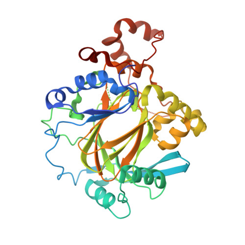
    5F2S, 5F2W, 5F32, 5F37, 5F39, 5F3C, 5F3E, 5F3G, 5F3I, 5F5A, 5F5C, 5F5I, 5FPL

  • PubMed Abstract: 

    We report the discovery of N-substituted 4-(pyridin-2-yl)thiazole-2-amine derivatives and their subsequent optimization, guided by structure-based design, to give 8-(1H-pyrazol-3-yl)pyrido[3,4-d]pyrimidin-4(3H)-ones, a series of potent JmjC histone N-methyl lysine demethylase (KDM) inhibitors which bind to Fe(II) in the active site. Substitution from C4 of the pyrazole moiety allows access to the histone peptide substrate binding site; incorporation of a conformationally constrained 4-phenylpiperidine linker gives derivatives such as 54j and 54k which demonstrate equipotent activity versus the KDM4 (JMJD2) and KDM5 (JARID1) subfamily demethylases, selectivity over representative exemplars of the KDM2, KDM3, and KDM6 subfamilies, cellular permeability in the Caco-2 assay, and, for 54k, inhibition of H3K9Me3 and H3K4Me3 demethylation in a cell-based assay.


  • Organizational Affiliation

    Cancer Research UK Cancer Therapeutics Unit, The Institute of Cancer Research , 15 Cotswold Road, London SM2 5NG, U.K.


Macromolecules
Find similar proteins by:  (by identity cutoff)  |  3D Structure
Entity ID: 1
MoleculeChains Sequence LengthOrganismDetailsImage
Lysine-specific demethylase 4A
A, B, C, D
360Homo sapiensMutation(s): 0 
Gene Names: KDM4AJHDM3AJMJD2JMJD2AKIAA0677
EC: 1.14.11
UniProt & NIH Common Fund Data Resources
Find proteins for O75164 (Homo sapiens)
Explore O75164 
Go to UniProtKB:  O75164
PHAROS:  O75164
GTEx:  ENSG00000066135 
Entity Groups  
Sequence Clusters30% Identity50% Identity70% Identity90% Identity95% Identity100% Identity
UniProt GroupO75164
Sequence Annotations
Expand
  • Reference Sequence
Small Molecules
Ligands 4 Unique
IDChains Name / Formula / InChI Key2D Diagram3D Interactions
5UO
Query on 5UO

Download Ideal Coordinates CCD File 
G [auth A],
K [auth B],
O [auth C],
T [auth D]
8-[4-[2-[4-(4-chlorophenyl)piperidin-1-yl]ethyl]pyrazol-1-yl]-3~{H}-pyrido[3,4-d]pyrimidin-4-one
C23 H23 Cl N6 O
SQDIBEJSOSAPGF-UHFFFAOYSA-N
GOL
Query on GOL

Download Ideal Coordinates CCD File 
L [auth B],
P [auth C]
GLYCEROL
C3 H8 O3
PEDCQBHIVMGVHV-UHFFFAOYSA-N
DMS
Query on DMS

Download Ideal Coordinates CCD File 
H [auth A],
Q [auth C]
DIMETHYL SULFOXIDE
C2 H6 O S
IAZDPXIOMUYVGZ-UHFFFAOYSA-N
ZN
Query on ZN

Download Ideal Coordinates CCD File 
E [auth A]
F [auth A]
I [auth B]
J [auth B]
M [auth C]
E [auth A],
F [auth A],
I [auth B],
J [auth B],
M [auth C],
N [auth C],
R [auth D],
S [auth D]
ZINC ION
Zn
PTFCDOFLOPIGGS-UHFFFAOYSA-N
Binding Affinity Annotations 
IDSourceBinding Affinity
5UO Binding MOAD:  5F3E IC50: 138 (nM) from 1 assay(s)
BindingDB:  5F3E IC50: 138 (nM) from 1 assay(s)
Experimental Data & Validation

Experimental Data

  • Method: X-RAY DIFFRACTION
  • Resolution: 2.16 Å
  • R-Value Free: 0.210 
  • R-Value Work: 0.162 
  • R-Value Observed: 0.165 
  • Space Group: P 1 21 1
Unit Cell:
Length ( Å )Angle ( ˚ )
a = 57.912α = 90
b = 101.626β = 99.41
c = 142.412γ = 90
Software Package:
Software NamePurpose
BUSTERrefinement
XDSdata reduction
Aimlessdata scaling
PHASERphasing

Structure Validation

View Full Validation Report


Ligand Structure Quality Assessment 


Entry History & Funding Information

Deposition Data


Funding OrganizationLocationGrant Number
Cancer Research UKUnited KingdomC309/A11566

Revision History  (Full details and data files)

  • Version 1.0: 2016-01-20
    Type: Initial release
  • Version 1.1: 2016-03-09
    Changes: Database references
  • Version 1.2: 2024-01-10
    Changes: Data collection, Database references, Derived calculations, Refinement description