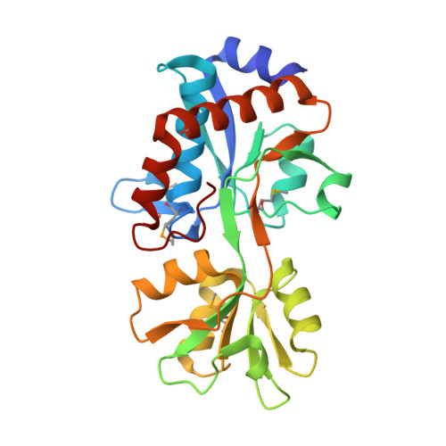
Crystal Structure of Solute-binding Protein from Enterococcus faecium with Bound Glutamate
Maltseva, N., Kim, Y., Mulligan, R., Shatsman, S., Anderson, W.F., Joachimiak, A., Center for Structural Genomics of Infectious Diseases (CSGID)To be published.