5EVY | pdb_00005evy

Salicylate hydroxylase substrate complex


Experimental Data Snapshot

  • Method: X-RAY DIFFRACTION
  • Resolution: 2.47 Å
  • R-Value Free: 
    0.260 (Depositor), 0.263 (DCC) 
  • R-Value Work: 
    0.201 (Depositor), 0.206 (DCC) 
  • R-Value Observed: 
    0.204 (Depositor) 

wwPDB Validation 3D Report Full Report

Validation slider image for 5EVY

Ligand Structure Quality Assessment 


This is version 1.3 of the entry. See complete history

Literature

The catalytic mechanism of decarboxylative hydroxylation of salicylate hydroxylase revealed by crystal structure analysis at 2.5 angstrom resolution

Uemura, T.Kita, A.Watanabe, Y.Adachi, M.Kuroki, R.Morimoto, Y.

(2016) Biochem Biophys Res Commun 469: 158-163

  • DOI: https://doi.org/10.1016/j.bbrc.2015.11.087
  • Primary Citation Related Structures: 
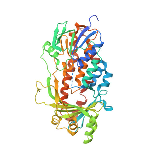
    5EVY

  • PubMed Abstract: 

    The X-ray crystal structure of a salicylate hydroxylase from Pseudomonas putida S-1 complexed with coenzyme FAD has been determined to a resolution of 2.5 Å. Structural conservation with p- or m-hydroxybenzoate hydroxylase is very good throughout the topology, despite a low amino sequence identity of 20-40% between these three hydroxylases. Salicylate hydroxylase is composed of three distinct domains and includes FAD between domains I and II, which is accessible to solvent. In this study, which analyzes the tertiary structure of the enzyme, the unique reaction of salicylate, i.e. decarboxylative hydroxylation, and the structural roles of amino acids surrounding the substrate, are considered.


  • Organizational Affiliation
    • Graduate School of Science, Kyoto University, Kyoto 606-8502, Japan; Research Reactor Institute, Kyoto University, Kumatori, Osaka 590-0494, Japan.

Macromolecule Content 

  • Total Structure Weight: 48.12 kDa 
  • Atom Count: 3,240 
  • Modeled Residue Count: 411 
  • Deposited Residue Count: 438 
  • Unique protein chains: 1

Macromolecules

Find similar proteins by:|  3D Structure
Entity ID: 1
MoleculeChains  Sequence LengthOrganismDetailsImage
Salicylate hydroxylaseA [auth X]438Pseudomonas putidaMutation(s): 0 
Gene Names: sal
EC: 1.14.13.1
UniProt
Find proteins for Q59713 (Pseudomonas putida)
Explore Q59713 
Go to UniProtKB:  Q59713
Entity Groups
Sequence Clusters30% Identity50% Identity70% Identity90% Identity95% Identity100% Identity
UniProt GroupQ59713
Sequence Annotations
Expand
Reference Sequence

Small Molecules

Ligands 2 Unique
IDChains Name / Formula / InChI Key2D Diagram3D Interactions
FAD

Query on FAD


Download:Ideal Coordinates CCD File
B [auth X]FLAVIN-ADENINE DINUCLEOTIDE
C27 H33 N9 O15 P2
VWWQXMAJTJZDQX-UYBVJOGSSA-N
SAL

Query on SAL


Download:Ideal Coordinates CCD File
C [auth X]2-HYDROXYBENZOIC ACID
C7 H6 O3
YGSDEFSMJLZEOE-UHFFFAOYSA-N

Experimental Data & Validation

Experimental Data

  • Method: X-RAY DIFFRACTION
  • Resolution: 2.47 Å
  • R-Value Free:  0.260 (Depositor), 0.263 (DCC) 
  • R-Value Work:  0.201 (Depositor), 0.206 (DCC) 
  • R-Value Observed: 0.204 (Depositor) 
Space Group: P 64
Unit Cell:
Length ( Å )Angle ( ˚ )
a = 142.33α = 90
b = 142.33β = 90
c = 62.35γ = 120
Software Package:
Software NamePurpose
REFMACrefinement
Cootmodel building
MOSFLMdata collection
SCALAdata scaling

Structure Validation

View Full Validation Report


Ligand Structure Quality Assessment 


Entry History 

Deposition Data

Revision History  (Full details and data files)

  • Version 1.0: 2015-12-16
    Type: Initial release
  • Version 1.1: 2016-01-13
    Changes: Database references
  • Version 1.2: 2020-02-19
    Changes: Data collection, Database references, Derived calculations
  • Version 1.3: 2024-03-20
    Changes: Data collection, Database references