Structural studies of a cold-adapted dimeric beta-D-galactosidase from Paracoccus sp. 32d.
Rutkiewicz-Krotewicz, M., Pietrzyk-Brzezinska, A.J., Sekula, B., Cieslinski, H., Wierzbicka-Wos, A., Kur, J., Bujacz, A.(2016) Acta Crystallogr D Struct Biol 72: 1049-1061
- PubMed: 27599737 Search on PubMed
- DOI: https://doi.org/10.1107/S2059798316012535
- Primary Citation Related Structures:
5EUV, 5LDR - PubMed Abstract:
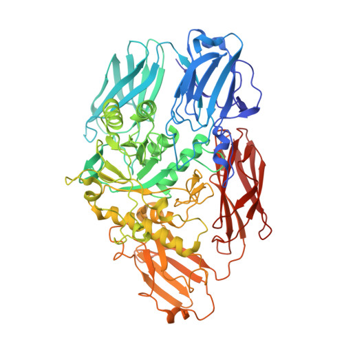
The crystal structure of a novel dimeric β-D-galactosidase from Paracoccus sp. 32d (ParβDG) was solved in space group P212121 at a resolution of 2.4 Å by molecular replacement with multiple models using the BALBES software. This enzyme belongs to glycoside hydrolase family 2 (GH2), similar to the tetrameric and hexameric β-D-galactosidases from Escherichia coli and Arthrobacter sp. C2-2, respectively. It is the second known structure of a cold-active GH2 β-galactosidase, and the first in the form of a functional dimer, which is also present in the asymmetric unit. Cold-adapted β-D-galactosidases have been the focus of extensive research owing to their utility in a variety of industrial technologies. One of their most appealing applications is in the hydrolysis of lactose, which not only results in the production of lactose-free dairy, but also eliminates the `sandy effect' and increases the sweetness of the product, thus enhancing its quality. The determined crystal structure represents the five-domain architecture of the enzyme, with its active site located in close vicinity to the dimer interface. To identify the amino-acid residues involved in the catalytic reaction and to obtain a better understanding of the mechanism of action of this atypical β-D-galactosidase, the crystal structure in complex with galactose (ParβDG-Gal) was also determined. The catalytic site of the enzyme is created by amino-acid residues from the central domain 3 and from domain 4 of an adjacent monomer. The crystal structure of this dimeric β-D-galactosidase reveals significant differences in comparison to other β-galactosidases. The largest difference is in the fifth domain, named Bgal_windup domain 5 in ParβDG, which contributes to stabilization of the functional dimer. The location of this domain 5, which is unique in size and structure, may be one of the factors responsible for the creation of a functional dimer and cold-adaptation of this enzyme.
- Institute of Technical Biochemistry, Faculty of Biotechnology and Food Sciences, Technical University of Lodz, Stefanowskiego 4/10, 90-924 Lodz, Poland.
Organizational Affiliation: