


A novel extracellular ligand receptor
Joachimiak, A.To be published.
Experimental Data Snapshot
wwPDB Validation 3D Report Full Report

Entity ID: 1 | |||||
|---|---|---|---|---|---|
| Molecule | Chains | Sequence Length | Organism | Details | Image |
| Extracellular ligand-binding receptor | 568 | Desulfohalobium retbaense DSM 5692 | Mutation(s): 0 Gene Names: Dret_0059 | 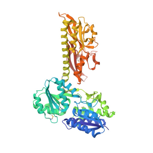 | |
UniProt | |||||
Entity Groups | |||||
| Sequence Clusters | 30% Identity50% Identity70% Identity90% Identity95% Identity100% Identity | ||||
| UniProt Group | C8WZ86 | ||||
Sequence AnnotationsExpand | |||||
Reference Sequence | |||||
| Ligands 6 Unique | |||||
|---|---|---|---|---|---|
| ID | Chains | Name / Formula / InChI Key | 2D Diagram | 3D Interactions | |
| COI Download:Ideal Coordinates CCD File | B [auth A] | 2-OXO-4-METHYLPENTANOIC ACID C6 H10 O3 BKAJNAXTPSGJCU-UHFFFAOYSA-N |  | ||
| CYT Download:Ideal Coordinates CCD File | C [auth A] | 6-AMINOPYRIMIDIN-2(1H)-ONE C4 H5 N3 O OPTASPLRGRRNAP-UHFFFAOYSA-N |  | ||
| GOL Download:Ideal Coordinates CCD File | F [auth A] | GLYCEROL C3 H8 O3 PEDCQBHIVMGVHV-UHFFFAOYSA-N |  | ||
| EDO Download:Ideal Coordinates CCD File | G [auth A], H [auth A] | 1,2-ETHANEDIOL C2 H6 O2 LYCAIKOWRPUZTN-UHFFFAOYSA-N |  | ||
| ACY Download:Ideal Coordinates CCD File | D [auth A] | ACETIC ACID C2 H4 O2 QTBSBXVTEAMEQO-UHFFFAOYSA-N |  | ||
| CA Download:Ideal Coordinates CCD File | E [auth A] | CALCIUM ION Ca BHPQYMZQTOCNFJ-UHFFFAOYSA-N |  | ||
| Modified Residues 1 Unique | |||||
|---|---|---|---|---|---|
| ID | Chains | Type | Formula | 2D Diagram | Parent |
| MSE Query on MSE | A | L-PEPTIDE LINKING | C5 H11 N O2 Se |  | MET |
| Length ( Å ) | Angle ( ˚ ) |
|---|---|
| a = 104.946 | α = 90 |
| b = 104.946 | β = 90 |
| c = 140.181 | γ = 90 |
| Software Name | Purpose |
|---|---|
| PHENIX | refinement |
| HKL-3000 | phasing |
| HKL-3000 | data reduction |


