5EQB

Crystal structure of lanosterol 14-alpha demethylase with intact transmembrane domain bound to itraconazole


Experimental Data Snapshot

  • Method: X-RAY DIFFRACTION
  • Resolution: 2.19 Å
  • R-Value Free: 0.228 
  • R-Value Work: 0.192 
  • R-Value Observed: 0.193 

wwPDB Validation   3D Report Full Report


Ligand Structure Quality Assessment 


This is version 1.1 of the entry. See complete history


Literature

Architecture of a single membrane spanning cytochrome P450 suggests constraints that orient the catalytic domain relative to a bilayer.

Monk, B.C.Tomasiak, T.M.Keniya, M.V.Huschmann, F.U.Tyndall, J.D.O'Connell, J.D.Cannon, R.D.McDonald, J.G.Rodriguez, A.Finer-Moore, J.S.Stroud, R.M.

(2014) Proc Natl Acad Sci U S A 111: 3865-3870

  • DOI: https://doi.org/10.1073/pnas.1324245111
  • Primary Citation of Related Structures:  
    4LXJ, 5EQB

  • PubMed Abstract: 

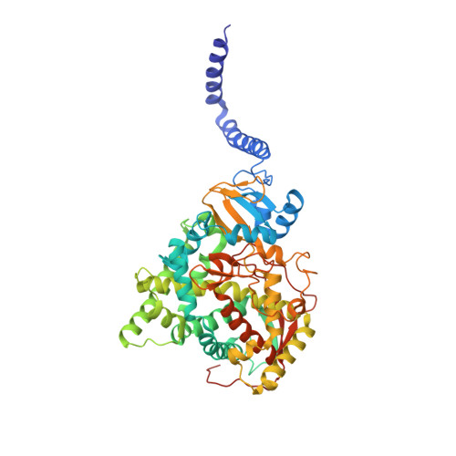
    Bitopic integral membrane proteins with a single transmembrane helix play diverse roles in catalysis, cell signaling, and morphogenesis. Complete monospanning protein structures are needed to show how interaction between the transmembrane helix and catalytic domain might influence association with the membrane and function. We report crystal structures of full-length Saccharomyces cerevisiae lanosterol 14α-demethylase, a membrane monospanning cytochrome P450 of the CYP51 family that catalyzes the first postcyclization step in ergosterol biosynthesis and is inhibited by triazole drugs. The structures reveal a well-ordered N-terminal amphipathic helix preceding a putative transmembrane helix that would constrain the catalytic domain orientation to lie partly in the lipid bilayer. The structures locate the substrate lanosterol, identify putative substrate and product channels, and reveal constrained interactions with triazole antifungal drugs that are important for drug design and understanding drug resistance.


  • Organizational Affiliation

    Sir John Walsh Research Institute, Department of Oral Sciences, Faculty of Dentistry, and School of Pharmacy, University of Otago, Dunedin 9054, New Zealand.


Macromolecules
Find similar proteins by:  (by identity cutoff)  |  3D Structure
Entity ID: 1
MoleculeChains Sequence LengthOrganismDetailsImage
Lanosterol 14-alpha demethylase539Saccharomyces cerevisiae S288CMutation(s): 0 
Gene Names: ERG11CYP51YHR007C
EC: 1.14.13.70
Membrane Entity: Yes 
UniProt
Find proteins for P10614 (Saccharomyces cerevisiae (strain ATCC 204508 / S288c))
Explore P10614 
Go to UniProtKB:  P10614
Entity Groups  
Sequence Clusters30% Identity50% Identity70% Identity90% Identity95% Identity100% Identity
UniProt GroupP10614
Sequence Annotations
Expand
  • Reference Sequence
Small Molecules
Ligands 2 Unique
IDChains Name / Formula / InChI Key2D Diagram3D Interactions
1YN
Query on 1YN

Download Ideal Coordinates CCD File 
C [auth A]2-[(2R)-butan-2-yl]-4-{4-[4-(4-{[(2R,4S)-2-(2,4-dichlorophenyl)-2-(1H-1,2,4-triazol-1-ylmethyl)-1,3-dioxolan-4-yl]methoxy}phenyl)piperazin-1-yl]phenyl}-2,4-dihydro-3H-1,2,4-triazol-3-one
C35 H38 Cl2 N8 O4
VHVPQPYKVGDNFY-DFMJLFEVSA-N
HEM
Query on HEM

Download Ideal Coordinates CCD File 
B [auth A]PROTOPORPHYRIN IX CONTAINING FE
C34 H32 Fe N4 O4
KABFMIBPWCXCRK-RGGAHWMASA-L
Experimental Data & Validation

Experimental Data

  • Method: X-RAY DIFFRACTION
  • Resolution: 2.19 Å
  • R-Value Free: 0.228 
  • R-Value Work: 0.192 
  • R-Value Observed: 0.193 
  • Space Group: P 1 21 1
Unit Cell:
Length ( Å )Angle ( ˚ )
a = 79.63α = 90
b = 67.845β = 99.75
c = 80.705γ = 90
Software Package:
Software NamePurpose
PHENIXrefinement
HKL-2000data reduction
HKL-2000data scaling
PHASERphasing

Structure Validation

View Full Validation Report


Ligand Structure Quality Assessment 


Entry History 

Revision History  (Full details and data files)

  • Version 1.0: 2016-01-13
    Type: Initial release
  • Version 1.1: 2024-03-06
    Changes: Data collection, Database references, Derived calculations