5EPH

Crystal structure of extended-spectrum beta-lactamase BEL-1 in complex with Imipenem


Experimental Data Snapshot

  • Method: X-RAY DIFFRACTION
  • Resolution: 1.79 Å
  • R-Value Free: 0.202 
  • R-Value Work: 0.164 
  • R-Value Observed: 0.165 

wwPDB Validation   3D Report Full Report


Ligand Structure Quality Assessment 


This is version 1.4 of the entry. See complete history


Literature

Crystal Structure of the Pseudomonas aeruginosa BEL-1 Extended-Spectrum beta-Lactamase and Its Complexes with Moxalactam and Imipenem.

Pozzi, C.De Luca, F.Benvenuti, M.Poirel, L.Nordmann, P.Rossolini, G.M.Mangani, S.Docquier, J.D.

(2016) Antimicrob Agents Chemother 60: 7189-7199

  • DOI: https://doi.org/10.1128/AAC.00936-16
  • Primary Citation of Related Structures:  
    5EOE, 5EOO, 5EPH, 5EUA

  • PubMed Abstract: 

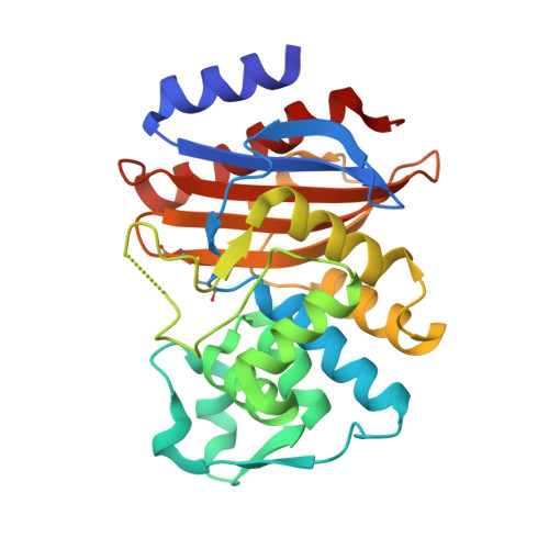
    BEL-1 is an acquired class A extended-spectrum β-lactamase (ESBL) found in Pseudomonas aeruginosa clinical isolates from Belgium which is divergent from other ESBLs (maximum identity of 54% with GES-type enzymes). This enzyme is efficiently inhibited by clavulanate, imipenem, and moxalactam. Crystals of BEL-1 were obtained at pH 5.6, and the structure of native BEL-1 was determined from orthorhombic and monoclinic crystal forms at 1.60-Å and 1.48-Å resolution, respectively. By soaking native BEL-1 crystals, complexes with imipenem (monoclinic form, 1.79-Å resolution) and moxalactam (orthorhombic form, 1.85-Å resolution) were also obtained. In the acyl-enzyme complexes, imipenem and moxalactam differ by the position of the α-substituent and of the carbonyl oxygen (in or out of the oxyanion hole). More surprisingly, the Ω-loop, which includes the catalytically relevant residue Glu166, was found in different conformations in the various subunits, resulting in the Glu166 side chain being rotated out of the active site or even in displacement of its Cα atom up to approximately 10 Å. A BEL-1 variant showing the single Leu162Phe substitution (BEL-2) confers a higher level of resistance to CAZ, CTX, and FEP and shows significantly lower K m values than BEL-1, especially with oxyiminocephalosporins. BEL-1 Leu162 is located at the beginning of the Ω-loop and is surrounded by Phe72, Leu139, and Leu148 (contact distances, 3.5 to 3.9 Å). This small hydrophobic cavity could not reasonably accommodate the bulkier Phe162 found in BEL-2 without altering neighboring residues or the Ω-loop itself, thus likely causing an important alteration of the enzyme kinetic properties.


  • Organizational Affiliation

    Dipartimento di Biotecnologie, Chimica e Farmacia, Università di Siena, Siena, Italy.


Macromolecules
Find similar proteins by:  (by identity cutoff)  |  3D Structure
Entity ID: 1
MoleculeChains Sequence LengthOrganismDetailsImage
Beta-lactamase
A, B, C, D
265Pseudomonas aeruginosaMutation(s): 0 
Gene Names: bla(BEL1)blaBEL-1
EC: 3.5.2.6
UniProt
Find proteins for Q3SAW3 (Pseudomonas aeruginosa)
Explore Q3SAW3 
Go to UniProtKB:  Q3SAW3
Entity Groups  
Sequence Clusters30% Identity50% Identity70% Identity90% Identity95% Identity100% Identity
UniProt GroupQ3SAW3
Sequence Annotations
Expand
  • Reference Sequence
Small Molecules
Ligands 3 Unique
IDChains Name / Formula / InChI Key2D Diagram3D Interactions
ID1
Query on ID1

Download Ideal Coordinates CCD File 
G [auth A],
I [auth B],
L [auth C],
N [auth D]
Imipenem
C12 H19 N3 O4 S
NIJGFQHESQFJAG-ZYUZMQFOSA-N
MRD
Query on MRD

Download Ideal Coordinates CCD File 
J [auth B](4R)-2-METHYLPENTANE-2,4-DIOL
C6 H14 O2
SVTBMSDMJJWYQN-RXMQYKEDSA-N
CL
Query on CL

Download Ideal Coordinates CCD File 
E [auth A],
F [auth A],
H [auth B],
K [auth C],
M [auth D]
CHLORIDE ION
Cl
VEXZGXHMUGYJMC-UHFFFAOYSA-M
Experimental Data & Validation

Experimental Data

  • Method: X-RAY DIFFRACTION
  • Resolution: 1.79 Å
  • R-Value Free: 0.202 
  • R-Value Work: 0.164 
  • R-Value Observed: 0.165 
  • Space Group: P 1 21 1
Unit Cell:
Length ( Å )Angle ( ˚ )
a = 53.98α = 90
b = 95.86β = 91.97
c = 103.42γ = 90
Software Package:
Software NamePurpose
REFMACrefinement
MOLREPmodel building
SCALAdata scaling
iMOSFLMdata reduction

Structure Validation

View Full Validation Report


Ligand Structure Quality Assessment 


Entry History 

Deposition Data

Revision History  (Full details and data files)

  • Version 1.0: 2016-09-21
    Type: Initial release
  • Version 1.1: 2016-10-05
    Changes: Database references
  • Version 1.2: 2016-12-07
    Changes: Database references
  • Version 1.3: 2018-01-31
    Changes: Database references, Source and taxonomy
  • Version 1.4: 2024-01-10
    Changes: Data collection, Database references, Refinement description