5ENM | pdb_00005enm

Compound 10


Experimental Data Snapshot

  • Method: X-RAY DIFFRACTION
  • Resolution: 1.98 Å
  • R-Value Free: 
    0.284 (Depositor), 0.278 (DCC) 
  • R-Value Work: 
    0.255 (Depositor), 0.252 (DCC) 
  • R-Value Observed: 
    0.256 (Depositor) 

wwPDB Validation 3D Report Full Report

Validation slider image for 5ENM

Ligand Structure Quality Assessment 


This is version 1.2 of the entry. See complete history

Literature

Targeting the BACE1 Active Site Flap Leads to a Potent Inhibitor That Elicits Robust Brain A beta Reduction in Rodents.

Wu, Y.J.Guernon, J.Yang, F.Snyder, L.Shi, J.Mcclure, A.Rajamani, R.Park, H.Ng, A.Lewis, H.Chang, C.Camac, D.Toyn, J.H.Ahlijanian, M.K.Albright, C.F.Macor, J.E.Thompson, L.A.

(2016) ACS Med Chem Lett 7: 271-276

  • DOI: https://doi.org/10.1021/acsmedchemlett.5b00432
  • Primary Citation Related Structures: 
    5ENK, 5ENM

  • PubMed Abstract: 

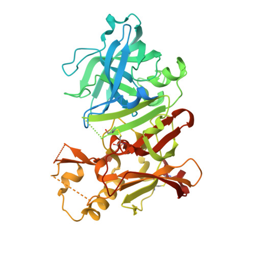
    By targeting the flap backbone of the BACE1 active site, we discovered 6-dimethylisoxazole-substituted biaryl aminothiazine 18 with 34-fold improved BACE1 inhibitory activity over the lead compound 1. The cocrystal structure of 18 bound to the active site indicated two hydrogen-bond interactions between the dimethylisoxazole and threonine 72 and glutamine 73 of the flap. Incorporation of the dimethylisoxazole substitution onto the related aminothiazine carboxamide series led to pyrazine-carboxamide 26 as a very potent BACE1 inhibitor (IC50 < 1 nM). This compound demonstrated robust brain Aβ reduction in rat dose-response studies. Thus, compound 26 may be useful in testing the amyloid hypothesis of Alzheimer's disease.


  • Organizational Affiliation
    • Research and Development, Bristol-Myers Squibb Company , 5 Research Parkway, Wallingford, Connecticut 06492-7660, United States.

Macromolecule Content 

  • Total Structure Weight: 50.84 kDa 
  • Atom Count: 2,889 
  • Modeled Residue Count: 359 
  • Deposited Residue Count: 455 
  • Unique protein chains: 1

Macromolecules

Find similar proteins by:|  3D Structure
Entity ID: 1
MoleculeChains  Sequence LengthOrganismDetailsImage
Beta-secretase 1455Homo sapiensMutation(s): 0 
Gene Names: BACE1BACEKIAA1149
EC: 3.4.23.46
UniProt & NIH Common Fund Data Resources
Find proteins for P56817 (Homo sapiens)
Explore P56817 
Go to UniProtKB:  P56817
PHAROS:  P56817
GTEx:  ENSG00000186318 
Entity Groups
Sequence Clusters30% Identity50% Identity70% Identity90% Identity95% Identity100% Identity
UniProt GroupP56817
Sequence Annotations
Expand
Reference Sequence

Small Molecules

Ligands 3 Unique
IDChains Name / Formula / InChI Key2D Diagram3D Interactions
5QU

Query on 5QU


Download:Ideal Coordinates CCD File
D [auth A](2~{R},4~{S},6~{S})-4-[2,4-bis(fluoranyl)-5-pyrimidin-5-yl-phenyl]-6-(3,5-dimethyl-1,2-oxazol-4-yl)-1,3-thiazinan-2-amine
C19 H19 F2 N5 O S
QXXCXVMLHLDXDN-JENIJYKNSA-N
IOD

Query on IOD


Download:Ideal Coordinates CCD File
C [auth A]IODIDE ION
I
XMBWDFGMSWQBCA-UHFFFAOYSA-M
GOL

Query on GOL


Download:Ideal Coordinates CCD File
B [auth A]GLYCEROL
C3 H8 O3
PEDCQBHIVMGVHV-UHFFFAOYSA-N

Experimental Data & Validation

Experimental Data

  • Method: X-RAY DIFFRACTION
  • Resolution: 1.98 Å
  • R-Value Free:  0.284 (Depositor), 0.278 (DCC) 
  • R-Value Work:  0.255 (Depositor), 0.252 (DCC) 
  • R-Value Observed: 0.256 (Depositor) 
Space Group: P 61 2 2
Unit Cell:
Length ( Å )Angle ( ˚ )
a = 102.234α = 90
b = 102.234β = 90
c = 170.579γ = 120
Software Package:
Software NamePurpose
REFMACrefinement
HKL-2000data reduction
autoPROCdata scaling
MOLREPphasing

Structure Validation

View Full Validation Report


Ligand Structure Quality Assessment 


Entry History 

Deposition Data

  • Released Date: 2016-10-05 
  • Deposition Author(s): Lewis, H.A.

Revision History  (Full details and data files)

  • Version 1.0: 2016-10-05
    Type: Initial release
  • Version 1.1: 2024-03-06
    Changes: Data collection, Database references, Derived calculations, Refinement description
  • Version 1.2: 2024-11-13
    Changes: Structure summary