5ELM | pdb_00005elm

Crystal structure of L-aspartate/glutamate specific racemase in complex with L-glutamate


Experimental Data Snapshot

  • Method: X-RAY DIFFRACTION
  • Resolution: 2.00 Å
  • R-Value Free: 
    0.236 (Depositor), 0.241 (DCC) 
  • R-Value Work: 
    0.173 (Depositor), 0.182 (DCC) 
  • R-Value Observed: 
    0.176 (Depositor) 

Starting Model: experimental
View more details

wwPDB Validation 3D Report Full Report

Validation slider image for 5ELM

This is version 1.2 of the entry. See complete history

Literature

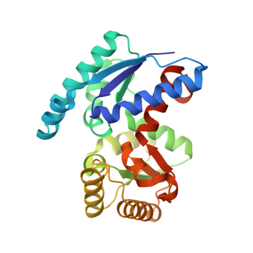
Structural basis for an atypical active site of an l-aspartate/glutamate-specific racemase from Escherichia coli

Ahn, J.W.Chang, J.H.Kim, K.J.

(2015) FEBS Lett 589: 3842-3847

  • DOI: https://doi.org/10.1016/j.febslet.2015.11.003
  • Primary Citation Related Structures: 
    5ELL, 5ELM

  • PubMed Abstract: 

    We determined the crystal structure of EcL-DER to elucidate protein function and substrate specificity. Unlike other asp/glu racemases, EcL-DER has an unbalanced pair of catalytic residues, Thr83/Cys197, at the active site that is crucial for L- to D-unidirectional racemase activity. EcL-DER exhibited racemase activity for both L-glutamate and L-aspartate, but had threefold higher activity for L-glutamate. Based on the structure of the EcL-DER(C197S) mutant in complex with L-glutamate, we determined the binding mode of the L-glutamate substrate in EcL-DER and provide a structural basis for how the protein utilizes L-glutamate as a main substrate. The unidirectionality, despite an equilibrium constant of unity, can be understood in terms of the Haldane relationship.


  • Organizational Affiliation
    • School of Life Sciences, KNU Creative BioResearch Group, Kyungpook National University, Daehak-ro 80, Buk-ku, Daegu 702-701, Republic of Korea.

Macromolecule Content 

  • Total Structure Weight: 106.24 kDa 
  • Atom Count: 7,990 
  • Modeled Residue Count: 940 
  • Deposited Residue Count: 952 
  • Unique protein chains: 1

Macromolecules

Find similar proteins by:|  3D Structure
Entity ID: 1
MoleculeChains  Sequence LengthOrganismDetailsImage
Asp/Glu_racemase family protein
A, B, C, D
238Escherichia coliMutation(s): 0 
EC: 5.1.1.13 (PDB Primary Data), 5.1.1 (UniProt), 5.1.1.10 (UniProt)
UniProt
Find proteins for C3SWD2 (Escherichia coli)
Explore C3SWD2 
Go to UniProtKB:  C3SWD2
Entity Groups
Sequence Clusters30% Identity50% Identity70% Identity90% Identity95% Identity100% Identity
UniProt GroupC3SWD2
Sequence Annotations
Expand
Reference Sequence

Experimental Data & Validation

Experimental Data

  • Method: X-RAY DIFFRACTION
  • Resolution: 2.00 Å
  • R-Value Free:  0.236 (Depositor), 0.241 (DCC) 
  • R-Value Work:  0.173 (Depositor), 0.182 (DCC) 
  • R-Value Observed: 0.176 (Depositor) 
Space Group: P 1 21 1
Unit Cell:
Length ( Å )Angle ( ˚ )
a = 67.53α = 90
b = 81.25β = 111.39
c = 83.96γ = 90
Software Package:
Software NamePurpose
REFMACrefinement
iMOSFLMdata reduction
SCALAdata scaling
MOLREPphasing

Structure Validation

View Full Validation Report


Entry History 

Deposition Data

Revision History  (Full details and data files)

  • Version 1.0: 2015-11-18
    Type: Initial release
  • Version 1.1: 2016-01-13
    Changes: Database references
  • Version 1.2: 2023-11-08
    Changes: Data collection, Database references, Derived calculations, Refinement description