Myosin MyTH4-FERM structures highlight important principles of convergent evolution.
Planelles-Herrero, V.J., Blanc, F., Sirigu, S., Sirkia, H., Clause, J., Sourigues, Y., Johnsrud, D.O., Amigues, B., Cecchini, M., Gilbert, S.P., Houdusse, A., Titus, M.A.(2016) Proc Natl Acad Sci U S A 113: E2906-E2915
- PubMed: 27166421 Search on PubMedSearch on PubMed Central
- DOI: https://doi.org/10.1073/pnas.1600736113
- Primary Citation Related Structures:
5EJQ, 5EJR, 5EJS, 5EJY - PubMed Abstract:
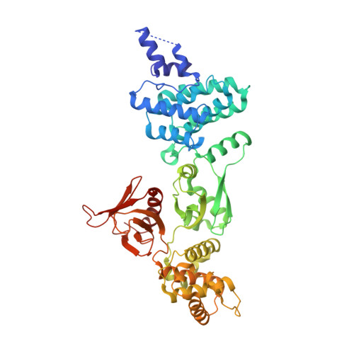
Myosins containing MyTH4-FERM (myosin tail homology 4-band 4.1, ezrin, radixin, moesin, or MF) domains in their tails are found in a wide range of phylogenetically divergent organisms, such as humans and the social amoeba Dictyostelium (Dd). Interestingly, evolutionarily distant MF myosins have similar roles in the extension of actin-filled membrane protrusions such as filopodia and bind to microtubules (MT), suggesting that the core functions of these MF myosins have been highly conserved over evolution. The structures of two DdMyo7 signature MF domains have been determined and comparison with mammalian MF structures reveals that characteristic features of MF domains are conserved. However, across millions of years of evolution conserved class-specific insertions are seen to alter the surfaces and the orientation of subdomains with respect to each other, likely resulting in new sites for binding partners. The MyTH4 domains of Myo10 and DdMyo7 bind to MT with micromolar affinity but, surprisingly, their MT binding sites are on opposite surfaces of the MyTH4 domain. The structural analysis in combination with comparison of diverse MF myosin sequences provides evidence that myosin tail domain features can be maintained without strict conservation of motifs. The results illustrate how tuning of existing features can give rise to new structures while preserving the general properties necessary for myosin tails. Thus, tinkering with the MF domain enables it to serve as a multifunctional platform for cooperative recruitment of various partners, allowing common properties such as autoinhibition of the motor and microtubule binding to arise through convergent evolution.
- Structural Motility, Institut Curie, CNRS, UMR 144, PSL Research University, F-75005 Paris, France; UPMC Université de Paris 6, Institut de Formation Doctorale, Sorbonne Universités, 75252 Paris Cedex 05, France;
Organizational Affiliation: