


Crystal structure of NAD kinase 1 from Listeria monocytogenes: V98S mutant
Poncet-Montange, G., Assairi, L., Gelin, M., Pochet, S., Labesse, G.To be published.
Experimental Data Snapshot
Starting Model: experimental
View more details
Entity ID: 1 | |||||
|---|---|---|---|---|---|
| Molecule | Chains | Sequence Length | Organism | Details | Image |
| NAD kinase 1 | 272 | Listeria monocytogenes | Mutation(s): 1 Gene Names: nadK1, lmo0968 EC: 2.7.1.23 | 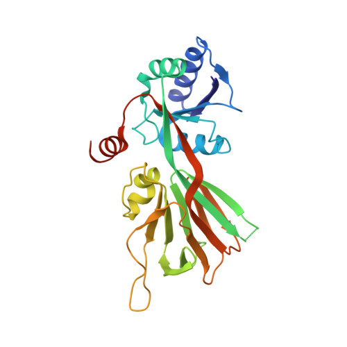 | |
UniProt | |||||
Entity Groups | |||||
| Sequence Clusters | 30% Identity50% Identity70% Identity90% Identity95% Identity100% Identity | ||||
| UniProt Group | Q8Y8D7 | ||||
Sequence AnnotationsExpand | |||||
Reference Sequence | |||||
| Ligands 1 Unique | |||||
|---|---|---|---|---|---|
| ID | Chains | Name / Formula / InChI Key | 2D Diagram | 3D Interactions | |
| NAP (Subject of Investigation/LOI) Download:Ideal Coordinates CCD File | B [auth A] | NADP NICOTINAMIDE-ADENINE-DINUCLEOTIDE PHOSPHATE C21 H28 N7 O17 P3 XJLXINKUBYWONI-NNYOXOHSSA-N |  | ||
| Length ( Å ) | Angle ( ˚ ) |
|---|---|
| a = 62.759 | α = 90 |
| b = 75.88 | β = 90 |
| c = 118.207 | γ = 90 |
| Software Name | Purpose |
|---|---|
| PHENIX | refinement |
| SCALA | data scaling |
| PDB_EXTRACT | data extraction |
| MOSFLM | data reduction |
| MOLREP | phasing |
| Funding Organization | Location | Grant Number |
|---|---|---|
| French National Research Agency | France | -- |


