5EH1 | pdb_00005eh1

Crystal structure of the extracellular part of receptor 2 of human interferon gamma


Experimental Data Snapshot

  • Method: X-RAY DIFFRACTION
  • Resolution: 1.80 Å
  • R-Value Free: 
    0.222 (Depositor), 0.189 (DCC) 
  • R-Value Work: 
    0.190 (Depositor) 
  • R-Value Observed: 
    0.191 (Depositor) 

wwPDB Validation 3D Report Full Report

Validation slider image for 5EH1

Ligand Structure Quality Assessment 


This is version 1.4 of the entry. See complete history

Literature

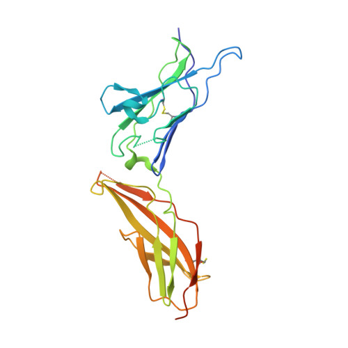
Crystal structure of human interferon-gamma receptor 2 reveals the structural basis for receptor specificity.

Mikulecky, P.Zahradnik, J.Kolenko, P.Cerny, J.Charnavets, T.Kolarova, L.Necasova, I.Pham, P.N.Schneider, B.

(2016) Acta Crystallogr D Struct Biol 72: 1017-1025

  • DOI: https://doi.org/10.1107/S2059798316012237
  • Primary Citation Related Structures: 
    5EH1

  • PubMed Abstract: 

    Interferon-γ receptor 2 is a cell-surface receptor that is required for interferon-γ signalling and therefore plays a critical immunoregulatory role in innate and adaptive immunity against viral and also bacterial and protozoal infections. A crystal structure of the extracellular part of human interferon-γ receptor 2 (IFNγR2) was solved by molecular replacement at 1.8 Å resolution. Similar to other class 2 receptors, IFNγR2 has two fibronectin type III domains. The characteristic structural features of IFNγR2 are concentrated in its N-terminal domain: an extensive π-cation motif of stacked residues KWRWRH, a NAG-W-NAG sandwich (where NAG stands for N-acetyl-D-glucosamine) and finally a helix formed by residues 78-85, which is unique among class 2 receptors. Mass spectrometry and mutational analyses showed the importance of N-linked glycosylation to the stability of the protein and confirmed the presence of two disulfide bonds. Structure-based bioinformatic analysis revealed independent evolutionary behaviour of both receptor domains and, together with multiple sequence alignment, identified putative binding sites for interferon-γ and receptor 1, the ligands of IFNγR2.


  • Organizational Affiliation
    • Institute of Biotechnology CAS, BIOCEV, Prumyslova 595, 252 50 Vestec, Czech Republic.

Macromolecule Content 

  • Total Structure Weight: 27.27 kDa 
  • Atom Count: 2,121 
  • Modeled Residue Count: 209 
  • Deposited Residue Count: 231 
  • Unique protein chains: 1

Macromolecules

Find similar proteins by:|  3D Structure
Entity ID: 1
MoleculeChains  Sequence LengthOrganismDetailsImage
Interferon gamma receptor 2231Homo sapiensMutation(s): 0 
Gene Names: IFNGR2IFNGT1
UniProt & NIH Common Fund Data Resources
Find proteins for P38484 (Homo sapiens)
Explore P38484 
Go to UniProtKB:  P38484
PHAROS:  P38484
GTEx:  ENSG00000159128 
Entity Groups
Sequence Clusters30% Identity50% Identity70% Identity90% Identity95% Identity100% Identity
UniProt GroupP38484
Glycosylation
Glycosylation Sites: 3Go to GlyGen: P38484-1
Sequence Annotations
Expand
Reference Sequence

Small Molecules

Ligands 3 Unique
IDChains Name / Formula / InChI Key2D Diagram3D Interactions
NAG

Query on NAG


Download:Ideal Coordinates CCD File
C [auth A],
D [auth A],
E [auth A]
2-acetamido-2-deoxy-beta-D-glucopyranose
C8 H15 N O6
OVRNDRQMDRJTHS-FMDGEEDCSA-N
CYS

Query on CYS


Download:Ideal Coordinates CCD File
B [auth A]CYSTEINE
C3 H7 N O2 S
XUJNEKJLAYXESH-REOHCLBHSA-N
GOL

Query on GOL


Download:Ideal Coordinates CCD File
F [auth A],
G [auth A],
H [auth A]
GLYCEROL
C3 H8 O3
PEDCQBHIVMGVHV-UHFFFAOYSA-N

Experimental Data & Validation

Experimental Data

  • Method: X-RAY DIFFRACTION
  • Resolution: 1.80 Å
  • R-Value Free:  0.222 (Depositor), 0.189 (DCC) 
  • R-Value Work:  0.190 (Depositor) 
  • R-Value Observed: 0.191 (Depositor) 
Space Group: P 61 2 2
Unit Cell:
Length ( Å )Angle ( ˚ )
a = 58.102α = 90
b = 58.102β = 90
c = 377.266γ = 120
Software Package:
Software NamePurpose
REFMACrefinement
MxCuBEdata collection
XDSdata reduction
BALBESphasing

Structure Validation

View Full Validation Report


Ligand Structure Quality Assessment 


Entry History 

Deposition Data

Revision History  (Full details and data files)

  • Version 1.0: 2016-08-17
    Type: Initial release
  • Version 1.1: 2016-09-14
    Changes: Database references
  • Version 1.2: 2018-04-18
    Changes: Data collection, Database references
  • Version 1.3: 2020-07-29
    Type: Remediation
    Reason: Carbohydrate remediation
    Changes: Data collection, Derived calculations, Structure summary
  • Version 1.4: 2024-11-20
    Changes: Data collection, Database references, Structure summary