5EGH | pdb_00005egh

Structure of ENPP6, a choline-specific glycerophosphodiester-phosphodiesterase in complex with phosphocholine


Experimental Data Snapshot

  • Method: X-RAY DIFFRACTION
  • Resolution: 1.80 Å
  • R-Value Free: 
    0.211 (Depositor), 0.214 (DCC) 
  • R-Value Work: 
    0.175 (Depositor), 0.177 (DCC) 
  • R-Value Observed: 
    0.177 (Depositor) 

wwPDB Validation 3D Report Full Report

Validation slider image for 5EGH

Ligand Structure Quality Assessment 


This is version 2.1 of the entry. See complete history

Literature

Structure and biological function of ENPP6, a choline-specific glycerophosphodiester-phosphodiesterase

Morita, J.Kano, K.Kato, K.Takita, H.Sakagami, H.Yamamoto, Y.Mihara, E.Ueda, H.Sato, T.Tokuyama, H.Arai, H.Asou, H.Takagi, J.Ishitani, R.Nishimasu, H.Nureki, O.Aoki, J.

(2016) Sci Rep 6: 20995-20995

  • DOI: https://doi.org/10.1038/srep20995
  • Primary Citation Related Structures: 
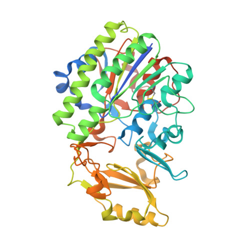
    5EGE, 5EGH

  • PubMed Abstract: 

    Choline is an essential nutrient for all living cells and is produced extracellularly by sequential degradation of phosphatidylcholine (PC). However, little is known about how choline is produced extracellularly. Here, we report that ENPP6, a choline-specific phosphodiesterase, hydrolyzes glycerophosphocholine (GPC), a degradation product of PC, as a physiological substrate and participates in choline metabolism. ENPP6 is highly expressed in liver sinusoidal endothelial cells and developing oligodendrocytes, which actively incorporate choline and synthesize PC. ENPP6-deficient mice exhibited fatty liver and hypomyelination, well known choline-deficient phenotypes. The choline moiety of GPC was incorporated into PC in an ENPP6-dependent manner both in vivo and in vitro. The crystal structure of ENPP6 in complex with phosphocholine revealed that the choline moiety of the phosphocholine is recognized by a choline-binding pocket formed by conserved aromatic and acidic residues. The present study provides the molecular basis for ENPP6-mediated choline metabolism at atomic, cellular and tissue levels.


  • Organizational Affiliation
    • Department of Biological Sciences, Graduate School of Sciences, The University of Tokyo, 2-11-16, Yayoi, Bunkyo-ku, Tokyo, 113-0032, Japan.

Macromolecule Content 

  • Total Structure Weight: 102.77 kDa 
  • Atom Count: 7,187 
  • Modeled Residue Count: 785 
  • Deposited Residue Count: 858 
  • Unique protein chains: 1

Macromolecules

Find similar proteins by:|  3D Structure
Entity ID: 1
MoleculeChains  Sequence LengthOrganismDetailsImage
Ectonucleotide pyrophosphatase/phosphodiesterase family member 6
A, B
429Mus musculusMutation(s): 2 
Gene Names: Enpp6
EC: 3.1.4 (PDB Primary Data), 3.1.4.38 (PDB Primary Data)
UniProt
Find proteins for Q8BGN3 (Mus musculus)
Explore Q8BGN3 
Go to UniProtKB:  Q8BGN3
Entity Groups
Sequence Clusters30% Identity50% Identity70% Identity90% Identity95% Identity100% Identity
UniProt GroupQ8BGN3
Glycosylation
Glycosylation Sites: 3Go to GlyGen: Q8BGN3-1
Sequence Annotations
Expand
Reference Sequence

Oligosaccharides

Help  
Entity ID: 2
MoleculeChains Length2D Diagram GlycosylationD Interactions
2-acetamido-2-deoxy-beta-D-glucopyranose-(1-4)-2-acetamido-2-deoxy-beta-D-glucopyranose
C, E, G
2N-Glycosylation
Glycosylation Resources
GlyTouCan: G42666HT
GlyCosmos: G42666HT
GlyGen: G42666HT
Entity ID: 3
MoleculeChains Length2D Diagram GlycosylationD Interactions
2-acetamido-2-deoxy-beta-D-glucopyranose-(1-4)-[beta-L-fucopyranose-(1-6)]2-acetamido-2-deoxy-beta-D-glucopyranose
D, F
3N-Glycosylation
Glycosylation Resources
GlyTouCan: G28454KX
GlyCosmos: G28454KX
GlyGen: G28454KX

Small Molecules

Ligands 4 Unique
IDChains Name / Formula / InChI Key2D Diagram3D Interactions
NAG

Query on NAG


Download:Ideal Coordinates CCD File
N [auth B]2-acetamido-2-deoxy-beta-D-glucopyranose
C8 H15 N O6
OVRNDRQMDRJTHS-FMDGEEDCSA-N
PC

Query on PC


Download:Ideal Coordinates CCD File
L [auth A],
R [auth B]
PHOSPHOCHOLINE
C5 H15 N O4 P
YHHSONZFOIEMCP-UHFFFAOYSA-O
ZN

Query on ZN


Download:Ideal Coordinates CCD File
H [auth A],
I [auth A],
O [auth B],
P [auth B]
ZINC ION
Zn
PTFCDOFLOPIGGS-UHFFFAOYSA-N
EDO

Query on EDO


Download:Ideal Coordinates CCD File
J [auth A],
K [auth A],
M [auth A],
Q [auth B],
S [auth B]
1,2-ETHANEDIOL
C2 H6 O2
LYCAIKOWRPUZTN-UHFFFAOYSA-N

Experimental Data & Validation

Experimental Data

  • Method: X-RAY DIFFRACTION
  • Resolution: 1.80 Å
  • R-Value Free:  0.211 (Depositor), 0.214 (DCC) 
  • R-Value Work:  0.175 (Depositor), 0.177 (DCC) 
  • R-Value Observed: 0.177 (Depositor) 
Space Group: P 1
Unit Cell:
Length ( Å )Angle ( ˚ )
a = 63.62α = 60.59
b = 68.849β = 86.99
c = 69.761γ = 68.1
Software Package:
Software NamePurpose
PHENIXrefinement
XSCALEdata scaling
MOLREPphasing
Cootmodel building
PDB_EXTRACTdata extraction

Structure Validation

View Full Validation Report


Ligand Structure Quality Assessment 


Entry History 

Deposition Data

Revision History  (Full details and data files)

  • Version 1.0: 2016-03-09
    Type: Initial release
  • Version 2.0: 2020-07-29
    Type: Remediation
    Reason: Carbohydrate remediation
    Changes: Atomic model, Data collection, Derived calculations, Structure summary
  • Version 2.1: 2024-10-23
    Changes: Data collection, Database references, Derived calculations, Structure summary