5EDJ | pdb_00005edj

Crystal structure of the Neisseria meningitidis iron-regulated outer membrane lipoprotein FrpD


Experimental Data Snapshot

  • Method: X-RAY DIFFRACTION
  • Resolution: 2.30 Å
  • R-Value Free: 
    0.249 (Depositor), 0.214 (DCC) 
  • R-Value Work: 
    0.183 (Depositor) 
  • R-Value Observed: 
    0.186 (Depositor) 

Starting Model: experimental
View more details

wwPDB Validation 3D Report Full Report

Validation slider image for 5EDJ

This is version 1.3 of the entry. See complete history

Literature

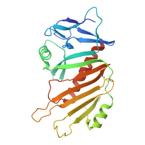
Structural basis of the interaction between the putative adhesion-involved and iron-regulated FrpD and FrpC proteins of Neisseria meningitidis.

Sviridova, E.Rezacova, P.Bondar, A.Veverka, V.Novak, P.Schenk, G.Svergun, D.I.Kuta Smatanova, I.Bumba, L.

(2017) Sci Rep 7: 40408-40408

  • DOI: https://doi.org/10.1038/srep40408
  • Primary Citation Related Structures: 
    5EDF, 5EDJ

  • PubMed Abstract: 

    The iron-regulated protein FrpD from Neisseria meningitidis is an outer membrane lipoprotein that interacts with very high affinity (K d  ~ 0.2 nM) with the N-terminal domain of FrpC, a Type I-secreted protein from the Repeat in ToXin (RTX) protein family. In the presence of Ca 2+ , FrpC undergoes Ca 2+ -dependent protein trans-splicing that includes an autocatalytic cleavage of the Asp 414 -Pro 415 peptide bond and formation of an Asp 414 -Lys isopeptide bond. Here, we report the high-resolution structure of FrpD and describe the structure-function relationships underlying the interaction between FrpD and FrpC 1-414 . We identified FrpD residues involved in FrpC 1-414 binding, which enabled localization of FrpD within the low-resolution SAXS model of the FrpD-FrpC 1-414 complex. Moreover, the trans-splicing activity of FrpC resulted in covalent linkage of the FrpC 1-414 fragment to plasma membrane proteins of epithelial cells in vitro, suggesting that formation of the FrpD-FrpC 1-414 complex may be involved in the interaction of meningococci with the host cell surface.


  • Organizational Affiliation
    • Faculty of Science, University of South Bohemia Ceske Budejovice, Branisovska 1760, 37005 Ceske Budejovice, Czech Republic.

Macromolecule Content 

  • Total Structure Weight: 27.03 kDa 
  • Atom Count: 1,979 
  • Modeled Residue Count: 224 
  • Deposited Residue Count: 231 
  • Unique protein chains: 1

Macromolecules

Find similar proteins by:|  3D Structure
Entity ID: 1
MoleculeChains  Sequence LengthOrganismDetailsImage
FrpC operon protein231Neisseria meningitidis MC58Mutation(s): 0 
Gene Names: LA50_03295
UniProt
Find proteins for Q9K129 (Neisseria meningitidis serogroup B (strain ATCC BAA-335 / MC58))
Explore Q9K129 
Go to UniProtKB:  Q9K129
Entity Groups
Sequence Clusters30% Identity50% Identity70% Identity90% Identity95% Identity100% Identity
UniProt GroupQ9K129
Sequence Annotations
Expand
Reference Sequence

Experimental Data & Validation

Experimental Data

  • Method: X-RAY DIFFRACTION
  • Resolution: 2.30 Å
  • R-Value Free:  0.249 (Depositor), 0.214 (DCC) 
  • R-Value Work:  0.183 (Depositor) 
  • R-Value Observed: 0.186 (Depositor) 
Space Group: P 64
Unit Cell:
Length ( Å )Angle ( ˚ )
a = 115.39α = 90
b = 115.39β = 90
c = 38.812γ = 120
Software Package:
Software NamePurpose
REFMACrefinement
SCALEPACKdata scaling
HKL-3000data reduction
MOLREPphasing

Structure Validation

View Full Validation Report


Entry History 

& Funding Information

Deposition Data


Funding OrganizationLocationGrant Number
Grant AgencyCzech RepublicP207/11/0717

Revision History  (Full details and data files)

  • Version 1.0: 2017-02-01
    Type: Initial release
  • Version 1.1: 2017-11-22
    Changes: Database references
  • Version 1.2: 2024-01-10
    Changes: Data collection, Database references, Refinement description
  • Version 1.3: 2024-11-20
    Changes: Structure summary