5E6Y

Crystal structure of E.Coli branching enzyme in complex with alpha cyclodextrin


Experimental Data Snapshot

  • Method: X-RAY DIFFRACTION
  • Resolution: 2.60 Å
  • R-Value Free: 0.256 
  • R-Value Work: 0.190 
  • R-Value Observed: 0.197 

wwPDB Validation   3D Report Full Report


This is version 2.1 of the entry. See complete history


Literature

Crystal structures of Escherichia coli branching enzyme in complex with cyclodextrins.

Feng, L.Fawaz, R.Hovde, S.Sheng, F.Nosrati, M.Geiger, J.H.

(2016) Acta Crystallogr D Struct Biol 72: 641-647

  • DOI: https://doi.org/10.1107/S2059798316003272
  • Primary Citation of Related Structures:  
    5E6Y, 5E6Z, 5E70

  • PubMed Abstract: 

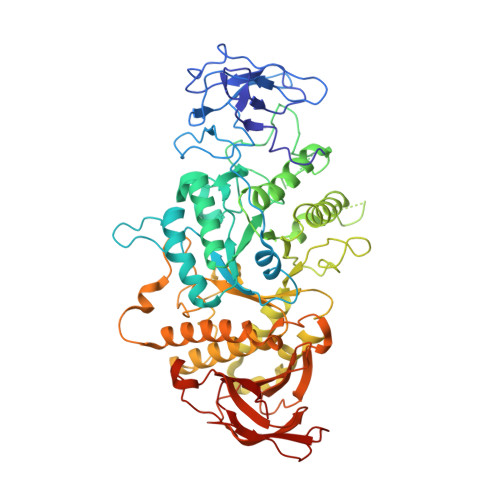
    Branching enzyme (BE) is responsible for the third step in glycogen/starch biosynthesis. It catalyzes the cleavage of α-1,4 glucan linkages and subsequent reattachment to form α-1,6 branch points. These branches are crucial to the final structure of glycogen and starch. The crystal structures of Escherichia coli BE (EcBE) in complex with α-, β- and γ-cyclodextrin were determined in order to better understand substrate binding. Four cyclodextrin-binding sites were identified in EcBE; they were all located on the surface of the enzyme, with none in the vicinity of the active site. While three of the sites were also identified as linear polysaccharide-binding sites, one of the sites is specific for cyclodextrins. In previous work three additional binding sites were identified as exclusively binding linear malto-oligosaccharides. Comparison of the binding sites shed light on this apparent specificity. Binding site IV is located in the carbohydrate-binding module 48 (CBM48) domain of EcBE and superimposes with the cyclodextrin-binding site found in the CBM48 domain of 5'-AMP-activated protein kinase (AMPK). Comparison of these sites shows the similarities and differences in the two binding modes. While some of the binding sites were found to be conserved between branching enzymes of different organisms, some are quite divergent, indicating both similarities and differences between oligosaccharide binding in branching enzymes from various sources.


  • Organizational Affiliation

    Department of Chemistry, Michigan State University, 578 South Shaw Lane, East Lansing, MI 48824, USA.


Macromolecules
Find similar proteins by:  (by identity cutoff)  |  3D Structure
Entity ID: 1
MoleculeChains Sequence LengthOrganismDetailsImage
1,4-alpha-glucan branching enzyme GlgB
A, B, C, D
612Escherichia coli O139:H28 str. E24377AMutation(s): 0 
Gene Names: glgBEcE24377A_3911
EC: 2.4.1.18
UniProt
Find proteins for A7ZSW5 (Escherichia coli O139:H28 (strain E24377A / ETEC))
Explore A7ZSW5 
Go to UniProtKB:  A7ZSW5
Entity Groups  
Sequence Clusters30% Identity50% Identity70% Identity90% Identity95% Identity100% Identity
UniProt GroupA7ZSW5
Sequence Annotations
Expand
  • Reference Sequence
Oligosaccharides

Help

Entity ID: 2
MoleculeChains Length2D Diagram Glycosylation3D Interactions
Cyclohexakis-(1-4)-(alpha-D-glucopyranose)
E, F, G, H, I
E, F, G, H, I, J, K, L, M
6N/A
Glycosylation Resources
GlyTouCan:  G22307HL
GlyCosmos:  G22307HL
Experimental Data & Validation

Experimental Data

  • Method: X-RAY DIFFRACTION
  • Resolution: 2.60 Å
  • R-Value Free: 0.256 
  • R-Value Work: 0.190 
  • R-Value Observed: 0.197 
  • Space Group: P 1 21 1
Unit Cell:
Length ( Å )Angle ( ˚ )
a = 92.024α = 90
b = 103.808β = 91.97
c = 186.08γ = 90
Software Package:
Software NamePurpose
PHENIXrefinement
HKL-2000data reduction
HKL-2000data scaling
MOLREPphasing

Structure Validation

View Full Validation Report


Entry History 

Deposition Data

Revision History  (Full details and data files)

  • Version 1.0: 2015-12-16
    Type: Initial release
  • Version 1.1: 2016-05-11
    Changes: Database references
  • Version 2.0: 2020-07-29
    Type: Remediation
    Reason: Carbohydrate remediation
    Changes: Atomic model, Data collection, Derived calculations, Non-polymer description, Structure summary
  • Version 2.1: 2023-09-27
    Changes: Data collection, Database references, Refinement description, Structure summary