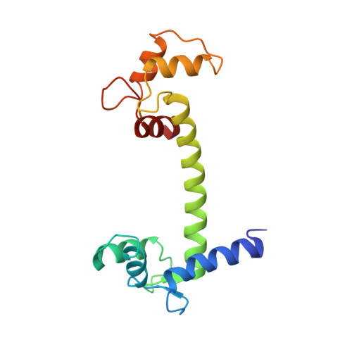
Atomic resolution experimental phase information reveals extensive disorder and bound 2-methyl-2,4-pentanediol in Ca(2+)-calmodulin.
Lin, J., van den Bedem, H., Brunger, A.T., Wilson, M.A.(2016) Acta Crystallogr D Struct Biol 72: 83-92
- PubMed: 26894537 Search on PubMedSearch on PubMed Central
- DOI: https://doi.org/10.1107/S2059798315021609
- Primary Citation Related Structures:
5E1K, 5E1N, 5E1P - PubMed Abstract:
Calmodulin (CaM) is the primary calcium signaling protein in eukaryotes and has been extensively studied using various biophysical techniques. Prior crystal structures have noted the presence of ambiguous electron density in both hydrophobic binding pockets of Ca(2+)-CaM, but no assignment of these features has been made. In addition, Ca(2+)-CaM samples many conformational substates in the crystal and accurately modeling the full range of this functionally important disorder is challenging. In order to characterize these features in a minimally biased manner, a 1.0 Å resolution single-wavelength anomalous diffraction data set was measured for selenomethionine-substituted Ca(2+)-CaM. Density-modified electron-density maps enabled the accurate assignment of Ca(2+)-CaM main-chain and side-chain disorder. These experimental maps also substantiate complex disorder models that were automatically built using low-contour features of model-phased electron density. Furthermore, experimental electron-density maps reveal that 2-methyl-2,4-pentanediol (MPD) is present in the C-terminal domain, mediates a lattice contact between N-terminal domains and may occupy the N-terminal binding pocket. The majority of the crystal structures of target-free Ca(2+)-CaM have been derived from crystals grown using MPD as a precipitant, and thus MPD is likely to be bound in functionally critical regions of Ca(2+)-CaM in most of these structures. The adventitious binding of MPD helps to explain differences between the Ca(2+)-CaM crystal and solution structures and is likely to favor more open conformations of the EF-hands in the crystal.
- Department of Biochemistry and Redox Biology Center, University of Nebraska, Beadle Center, Lincoln, NE 68588, USA.
Organizational Affiliation: