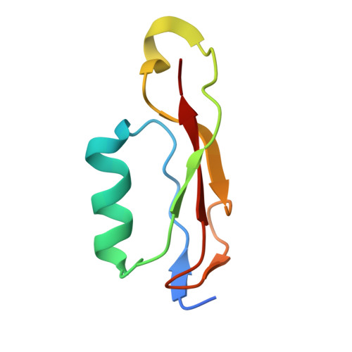
Structural and Genetic Analyses of the Mycobacterium tuberculosis Protein Kinase B Sensor Domain Identify a Potential Ligand-binding Site.
Prigozhin, D.M., Papavinasasundaram, K.G., Baer, C.E., Murphy, K.C., Moskaleva, A., Chen, T.Y., Alber, T., Sassetti, C.M.(2016) J Biological Chem 291: 22961-22969
- PubMed: 27601474 Search on PubMedSearch on PubMed Central
- DOI: https://doi.org/10.1074/jbc.M116.731760
- Primary Citation Related Structures:
3OUV, 5E0Y, 5E0Z, 5E10, 5E12 - PubMed Abstract:
Monitoring the environment with serine/threonine protein kinases is critical for growth and survival of Mycobacterium tuberculosis, a devastating human pathogen. Protein kinase B (PknB) is a transmembrane serine/threonine protein kinase that acts as an essential regulator of mycobacterial growth and division. The PknB extracellular domain (ECD) consists of four repeats homologous to penicillin-binding protein and serine/threonine kinase associated (PASTA) domains, and binds fragments of peptidoglycan. These properties suggest that PknB activity is modulated by ECD binding to peptidoglycan substructures, however, the molecular mechanisms underpinning PknB regulation remain unclear. In this study, we report structural and genetic characterization of the PknB ECD. We determined the crystal structures of overlapping ECD fragments at near atomic resolution, built a model of the full ECD, and discovered a region on the C-terminal PASTA domain that has the properties of a ligand-binding site. Hydrophobic interaction between this surface and a bound molecule of citrate was observed in a crystal structure. Our genetic analyses in M. tuberculosis showed that nonfunctional alleles were produced either by deletion of any of single PASTA domain or by mutation of individual conserved residues lining the putative ligand-binding surface of the C-terminal PASTA repeat. These results define two distinct structural features necessary for PknB signal transduction, a fully extended ECD and a conserved, membrane-distal putative ligand-binding site.
- From the Department of Molecular and Cell Biology, QB3 Institute, University of California, Berkeley, California 94720-3220 and.
Organizational Affiliation: