


Crystal structure of mouse CTLA-4 nanobody
Fedorov, A.A., Fedorov, E.V., Samanta, D., Bonanno, J.B., Almo, S.C.To be published.
Experimental Data Snapshot
Starting Model: experimental
View more details
wwPDB Validation 3D Report Full Report

Entity ID: 1 | |||||
|---|---|---|---|---|---|
| Molecule | Chains | Sequence Length | Organism | Details | Image |
| CTLA-4 nanobody | 129 | Camelidae | Mutation(s): 0 | 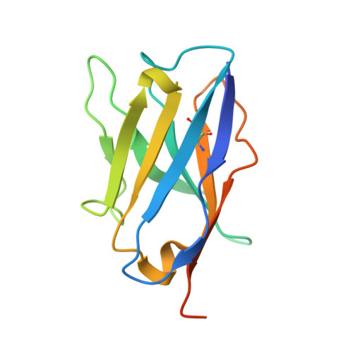 | |
Entity Groups | |||||
| Sequence Clusters | 30% Identity50% Identity70% Identity90% Identity95% Identity100% Identity | ||||
Sequence AnnotationsExpand | |||||
Reference Sequence | |||||
| Ligands 1 Unique | |||||
|---|---|---|---|---|---|
| ID | Chains | Name / Formula / InChI Key | 2D Diagram | 3D Interactions | |
| SO4 Download:Ideal Coordinates CCD File | B [auth A] | SULFATE ION O4 S QAOWNCQODCNURD-UHFFFAOYSA-L |  | ||
| Length ( Å ) | Angle ( ˚ ) |
|---|---|
| a = 42.147 | α = 90 |
| b = 42.147 | β = 90 |
| c = 208.517 | γ = 120 |
| Software Name | Purpose |
|---|---|
| PHENIX | refinement |
| CBASS | data collection |
| HKL-2000 | data scaling |
| PHASER | phasing |


