5DZZ | pdb_00005dzz

Structural characterization of intermediate filaments binding domain of desmoplakin


Experimental Data Snapshot

  • Method: X-RAY DIFFRACTION
  • Resolution: 2.60 Å
  • R-Value Free: 
    0.264 (Depositor), 0.266 (DCC) 
  • R-Value Work: 
    0.207 (Depositor), 0.211 (DCC) 
  • R-Value Observed: 
    0.211 (Depositor) 

Starting Model: experimental
View more details

wwPDB Validation 3D Report Full Report

Validation slider image for 5DZZ

This is version 1.2 of the entry. See complete history

Literature

Structure of the Intermediate Filament-Binding Region of Desmoplakin.

Kang, H.Weiss, T.M.Bang, I.Weis, W.I.Choi, H.J.

(2016) PLoS One 11: e0147641-e0147641

  • DOI: https://doi.org/10.1371/journal.pone.0147641
  • Primary Citation Related Structures: 
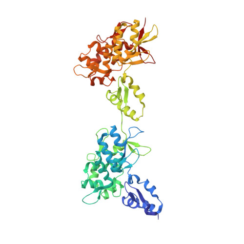
    5DZZ

  • PubMed Abstract: 

    Desmoplakin (DP) is a cytoskeletal linker protein that connects the desmosomal cadherin/plakoglobin/plakophilin complex to intermediate filaments (IFs). The C-terminal region of DP (DPCT) mediates IF binding, and contains three plakin repeat domains (PRDs), termed PRD-A, PRD-B and PRD-C. Previous crystal structures of PRDs B and C revealed that each is formed by 4.5 copies of a plakin repeat (PR) and has a conserved positively charged groove on its surface. Although PRDs A and B are linked by just four amino acids, B and C are separated by a 154 residue flexible linker, which has hindered crystallographic analysis of the full DPCT. Here we present the crystal structure of a DPCT fragment spanning PRDs A and B, and elucidate the overall architecture of DPCT by small angle X-ray scattering (SAXS) analysis. The structure of PRD-A is similar to that of PRD-B, and the two domains are arranged in a quasi-linear arrangement, and separated by a 4 amino acid linker. Analysis of the B-C linker region using secondary structure prediction and the crystal structure of a homologous linker from the cytolinker periplakin suggests that the N-terminal ~100 amino acids of the linker form two PR-like motifs. SAXS analysis of DPCT indicates an elongated but non-linear shape with Rg = 51.5 Å and Dmax = 178 Å. These data provide the first structural insights into an IF binding protein containing multiple PRDs and provide a foundation for studying the molecular basis of DP-IF interactions.


  • Organizational Affiliation
    • Dept. of Biological Sciences, Seoul National University, Seoul, South Korea.

Macromolecule Content 

  • Total Structure Weight: 54.56 kDa 
  • Atom Count: 3,884 
  • Modeled Residue Count: 489 
  • Deposited Residue Count: 494 
  • Unique protein chains: 1

Macromolecules

Find similar proteins by:|  3D Structure
Entity ID: 1
MoleculeChains  Sequence LengthOrganismDetailsImage
Desmoplakin494Homo sapiensMutation(s): 0 
Gene Names: DSP
UniProt & NIH Common Fund Data Resources
Find proteins for P15924 (Homo sapiens)
Explore P15924 
Go to UniProtKB:  P15924
PHAROS:  P15924
GTEx:  ENSG00000096696 
Entity Groups
Sequence Clusters30% Identity50% Identity70% Identity90% Identity95% Identity100% Identity
UniProt GroupP15924
Sequence Annotations
Expand
Reference Sequence

Experimental Data & Validation

Experimental Data

  • Method: X-RAY DIFFRACTION
  • Resolution: 2.60 Å
  • R-Value Free:  0.264 (Depositor), 0.266 (DCC) 
  • R-Value Work:  0.207 (Depositor), 0.211 (DCC) 
  • R-Value Observed: 0.211 (Depositor) 
Space Group: P 21 21 21
Unit Cell:
Length ( Å )Angle ( ˚ )
a = 111.931α = 90
b = 64.466β = 90
c = 74.044γ = 90
Software Package:
Software NamePurpose
SCALAdata scaling
Cootmodel building
PHENIXrefinement
PDB_EXTRACTdata extraction
SCALAdata reduction
PHASERphasing

Structure Validation

View Full Validation Report


Entry History 

Deposition Data

Revision History  (Full details and data files)

  • Version 1.0: 2016-03-23
    Type: Initial release
  • Version 1.1: 2019-12-04
    Changes: Data collection, Derived calculations
  • Version 1.2: 2023-11-08
    Changes: Data collection, Database references, Refinement description