5DYI | pdb_00005dyi

Structure of p97 N-D1 wild-type in complex with ADP


Experimental Data Snapshot

  • Method: X-RAY DIFFRACTION
  • Resolution: 3.71 Å
  • R-Value Free: 
    0.285 (Depositor), 0.282 (DCC) 
  • R-Value Work: 
    0.249 (Depositor), 0.249 (DCC) 
  • R-Value Observed: 
    0.250 (Depositor) 

wwPDB Validation 3D Report Full Report

Validation slider image for 5DYI

Ligand Structure Quality Assessment 


This is version 1.1 of the entry. See complete history

Literature

Role of the D1-D2 Linker of Human VCP/p97 in the Asymmetry and ATPase Activity of the D1-domain.

Tang, W.K.Xia, D.

(2016) Sci Rep 6: 20037-20037

  • DOI: https://doi.org/10.1038/srep20037
  • Primary Citation Related Structures: 
    5DYG, 5DYI

  • PubMed Abstract: 

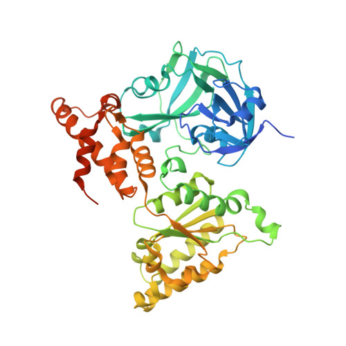
    Human AAA(+) protein p97 consists of an N-domain and two tandem ATPase domains D1 and D2, which are connected by the N-D1 and the D1-D2 linkers. Inclusion of the D1-D2 linker, a 22-amino acid peptide, at the end of p97 N-D1 truncate has been shown to activate ATP hydrolysis of its D1-domain, although the mechanism of activation remains unclear. Here, we identify the N-terminal half of this linker, highly conserved from human to fungi, is essential for the ATPase activation. By analyzing available crystal structures, we observed that the D1-D2 linker is capable of inducing asymmetry in subunit association into a p97 hexamer. This observation is reinforced by two new crystal structures, determined in the present work. The effect of D1-D2 linker on the ATPase activity of the D1-domain is correlated to the side-chain conformation of residue R359, a trans-acting arginine-finger residue essential for ATP hydrolysis of the D1-domain. The activation in D1-domain ATPase activity by breaking perfect six-fold symmetry implies functional importance of asymmetric association of p97 subunits, the extent of which can be determined quantitatively by the metric Asymmetric Index.


  • Organizational Affiliation
    • Laboratory of Cell Biology, Center for Cancer Research, National Cancer Institute, National Institutes of Health, Bethesda, MD 20892, USA.

Macromolecule Content 

  • Total Structure Weight: 660.1 kDa 
  • Atom Count: 41,579 
  • Modeled Residue Count: 5,265 
  • Deposited Residue Count: 5,868 
  • Unique protein chains: 1

Macromolecules

Find similar proteins by:|  3D Structure
Entity ID: 1
MoleculeChains  Sequence LengthOrganismDetailsImage
Transitional endoplasmic reticulum ATPase
A, B, C, D, E
A, B, C, D, E, F, G, H, I, J, K, L
489Homo sapiensMutation(s): 0 
Gene Names: VCP
EC: 3.6.4.6
UniProt & NIH Common Fund Data Resources
Find proteins for P55072 (Homo sapiens)
Explore P55072 
Go to UniProtKB:  P55072
PHAROS:  P55072
GTEx:  ENSG00000165280 
Entity Groups
Sequence Clusters30% Identity50% Identity70% Identity90% Identity95% Identity100% Identity
UniProt GroupP55072
Sequence Annotations
Expand
Reference Sequence

Small Molecules

Ligands 1 Unique
IDChains Name / Formula / InChI Key2D Diagram3D Interactions
ADP

Query on ADP


Download:Ideal Coordinates CCD File
M [auth A]
N [auth B]
O [auth C]
P [auth D]
Q [auth E]
M [auth A],
N [auth B],
O [auth C],
P [auth D],
Q [auth E],
R [auth F],
S [auth G],
T [auth H],
U [auth I],
V [auth J],
W [auth K],
X [auth L]
ADENOSINE-5'-DIPHOSPHATE
C10 H15 N5 O10 P2
XTWYTFMLZFPYCI-KQYNXXCUSA-N

Experimental Data & Validation

Experimental Data

  • Method: X-RAY DIFFRACTION
  • Resolution: 3.71 Å
  • R-Value Free:  0.285 (Depositor), 0.282 (DCC) 
  • R-Value Work:  0.249 (Depositor), 0.249 (DCC) 
  • R-Value Observed: 0.250 (Depositor) 
Space Group: P 21 21 21
Unit Cell:
Length ( Å )Angle ( ˚ )
a = 146.44α = 90
b = 176.21β = 90
c = 256.87γ = 90
Software Package:
Software NamePurpose
REFMACrefinement
DENZOdata reduction
SCALEPACKdata scaling
MOLREPphasing

Structure Validation

View Full Validation Report


Ligand Structure Quality Assessment 


Entry History 

Deposition Data

Revision History  (Full details and data files)

  • Version 1.0: 2016-02-10
    Type: Initial release
  • Version 1.1: 2024-03-06
    Changes: Data collection, Database references, Derived calculations, Refinement description