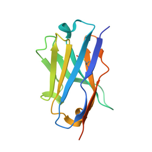
Crystal structure of mouse PD-L1 nanobody
Fedorov, A.A., Fedorov, E.V., Samanta, D., Almo, S.C.To be published.
Experimental Data Snapshot
Starting Model: experimental
View more details
wwPDB Validation 3D Report Full Report

| Ligands 1 Unique | |||||
|---|---|---|---|---|---|
| ID | Chains | Name / Formula / InChI Key | 2D Diagram | 3D Interactions | |
| SO4 Download:Ideal Coordinates CCD File | B [auth A], C [auth A] | SULFATE ION O4 S QAOWNCQODCNURD-UHFFFAOYSA-L |  | ||
| Length ( Å ) | Angle ( ˚ ) |
|---|---|
| a = 85.191 | α = 90 |
| b = 29.679 | β = 90 |
| c = 35.268 | γ = 90 |
| Software Name | Purpose |
|---|---|
| PHENIX | refinement |
| CBASS | data collection |
| HKL-2000 | data reduction |
| PHASER | phasing |
| HKL-2000 | data scaling |