5DX0

Crystal structure of CARM1, sinefungin, and H3 peptide (R17)


Experimental Data Snapshot

  • Method: X-RAY DIFFRACTION
  • Resolution: 2.05 Å
  • R-Value Free: 0.263 
  • R-Value Work: 0.211 
  • R-Value Observed: 0.214 

wwPDB Validation   3D Report Full Report


Ligand Structure Quality Assessment 


This is version 1.1 of the entry. See complete history


Literature

Structural Insights into Ternary Complex Formation of Human CARM1 with Various Substrates.

Boriack-Sjodin, P.A.Jin, L.Jacques, S.L.Drew, A.Sneeringer, C.Scott, M.P.Moyer, M.P.Ribich, S.Moradei, O.Copeland, R.A.

(2016) ACS Chem Biol 11: 763-771

  • DOI: https://doi.org/10.1021/acschembio.5b00773
  • Primary Citation of Related Structures:  
    5DWQ, 5DX0, 5DX1, 5DX8, 5DXA, 5DXJ

  • PubMed Abstract: 

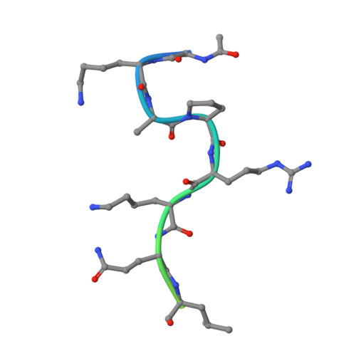
    Coactivator-associated arginine methyltransferase 1 (CARM1) is a protein arginine N-methyltransferase (PRMT) enzyme that has been implicated in a variety of cancers. CARM1 is known to methylate histone H3 and nonhistone substrates. To date, several crystal structures of CARM1 have been solved, including structures with small molecule inhibitors, but no ternary structures with nucleoside and peptide substrates have been reported. Here, the crystal structures of human CARM1 with the S-adenosylmethione (SAM) mimic sinefungin and three different peptide sequences from histone H3 and PABP1 are presented, with both nonmethylated and singly methylated arginine residues exemplified. This is the first example of multiple substrate sequences solved in a single PRMT enzyme and demonstrates how the CARM1 binding site is capable of accommodating a variety of peptide sequences while maintaining a core binding mode for the unmethylated and monomethylated substrates. Comparison of these with other PRMT enzyme-peptide structures shows hydrogen bonding patterns that may be thematic of these binding sites.


  • Organizational Affiliation

    Epizyme, Inc. 400 Technology Square, Cambridge, Massachusetts 02139, United States.


Macromolecules
Find similar proteins by:  (by identity cutoff)  |  3D Structure
Entity ID: 1
MoleculeChains Sequence LengthOrganismDetailsImage
Histone-arginine methyltransferase CARM1
A, B, C, D
349Homo sapiensMutation(s): 0 
Gene Names: CARM1PRMT4
EC: 2.1.1 (PDB Primary Data), 2.1.1.125 (PDB Primary Data)
UniProt & NIH Common Fund Data Resources
Find proteins for Q86X55 (Homo sapiens)
Explore Q86X55 
Go to UniProtKB:  Q86X55
PHAROS:  Q86X55
GTEx:  ENSG00000142453 
Entity Groups  
Sequence Clusters30% Identity50% Identity70% Identity90% Identity95% Identity100% Identity
UniProt GroupQ86X55
Sequence Annotations
Expand
  • Reference Sequence

Find similar proteins by:  Sequence   |   3D Structure  

Entity ID: 2
MoleculeChains Sequence LengthOrganismDetailsImage
H3 peptideE [auth F],
F [auth G],
G [auth H],
H [auth I]
20Homo sapiensMutation(s): 2 
UniProt & NIH Common Fund Data Resources
Find proteins for P84243 (Homo sapiens)
Explore P84243 
Go to UniProtKB:  P84243
PHAROS:  P84243
Entity Groups  
Sequence Clusters30% Identity50% Identity70% Identity90% Identity95% Identity100% Identity
UniProt GroupP84243
Sequence Annotations
Expand
  • Reference Sequence
Small Molecules
Ligands 3 Unique
IDChains Name / Formula / InChI Key2D Diagram3D Interactions
SFG
Query on SFG

Download Ideal Coordinates CCD File 
I [auth A],
L [auth B],
N [auth C],
Q [auth D]
SINEFUNGIN
C15 H23 N7 O5
LMXOHSDXUQEUSF-YECHIGJVSA-N
SO4
Query on SO4

Download Ideal Coordinates CCD File 
K [auth A],
M [auth B],
P [auth C],
R [auth D]
SULFATE ION
O4 S
QAOWNCQODCNURD-UHFFFAOYSA-L
GOL
Query on GOL

Download Ideal Coordinates CCD File 
J [auth A],
O [auth C]
GLYCEROL
C3 H8 O3
PEDCQBHIVMGVHV-UHFFFAOYSA-N
Experimental Data & Validation

Experimental Data

  • Method: X-RAY DIFFRACTION
  • Resolution: 2.05 Å
  • R-Value Free: 0.263 
  • R-Value Work: 0.211 
  • R-Value Observed: 0.214 
  • Space Group: P 21 21 2
Unit Cell:
Length ( Å )Angle ( ˚ )
a = 75.722α = 90
b = 98.847β = 90
c = 208.67γ = 90
Software Package:
Software NamePurpose
HKL-2000data reduction
SCALEPACKdata scaling
REFMACrefinement
PDB_EXTRACTdata extraction

Structure Validation

View Full Validation Report


Ligand Structure Quality Assessment 


Entry History 

Deposition Data

Revision History  (Full details and data files)

  • Version 1.0: 2015-11-25
    Type: Initial release
  • Version 1.1: 2016-03-30
    Changes: Database references